Compounds of the upper gastrointestinal tract induce rapid and efficient excystation of Entamoeba invadens
- PMID: 20018192
- PMCID: PMC2881592
- DOI: 10.1016/j.ijpara.2009.11.012
Compounds of the upper gastrointestinal tract induce rapid and efficient excystation of Entamoeba invadens
Abstract
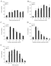
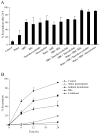
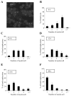
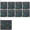
The infective stage of Entamoeba parasites is an encysted form. This stage can be readily generated in vitro, which has allowed identification of stimuli that trigger the differentiation of the parasite trophozoite stage into the cyst stage. Studies of the second differentiation event, emergence of the parasite from the cyst upon infection of a host, have been hampered by the lack of an efficient means to excyst the parasite and complete the life cycle in vitro. We have determined that a combination of exposures to water, bicarbonate and bile induces rapid excystment of Entamoeba invadens cysts. The high efficiency of this method has allowed the visualization of the dynamics of the process by electron and confocal microscopy, and should permit the analysis of stage-specific gene expression and high-throughput screening of inhibitory compounds.
(c) 2010 Australian Society for Parasitology Inc. Published by Elsevier Ltd. All rights reserved.
Figures


Similar articles
-
Entamoeba Chitinase is Required for Mature Round Cyst Formation.Microbiol Spectr. 2021 Sep 3;9(1):e0051121. doi: 10.1128/Spectrum.00511-21. Epub 2021 Aug 4. Microbiol Spectr. 2021. PMID: 34346756 Free PMC article.
-
A role for a galactose lectin and its ligands during encystment of Entamoeba.J Eukaryot Microbiol. 2001 Jan-Feb;48(1):17-21. doi: 10.1111/j.1550-7408.2001.tb00411.x. J Eukaryot Microbiol. 2001. PMID: 11249188 Review.
-
Identification of a developmentally regulated transcript expressed during encystation of Entamoeba invadens.Mol Biochem Parasitol. 1994 Sep;67(1):125-35. doi: 10.1016/0166-6851(94)90102-3. Mol Biochem Parasitol. 1994. PMID: 7838173
-
Eukaryotic Initiation Factor 2α Kinases Regulate Virulence Functions, Stage Conversion, and the Stress Response in Entamoeba invadens.mSphere. 2022 Jun 29;7(3):e0013122. doi: 10.1128/msphere.00131-22. Epub 2022 May 31. mSphere. 2022. PMID: 35638357 Free PMC article.
-
Encystation in parasitic protozoa.Curr Opin Microbiol. 2001 Aug;4(4):421-6. doi: 10.1016/s1369-5274(00)00229-0. Curr Opin Microbiol. 2001. PMID: 11495805 Review.
Cited by
-
Entamoeba Chitinase is Required for Mature Round Cyst Formation.Microbiol Spectr. 2021 Sep 3;9(1):e0051121. doi: 10.1128/Spectrum.00511-21. Epub 2021 Aug 4. Microbiol Spectr. 2021. PMID: 34346756 Free PMC article.
-
Characterization of Extracellular Vesicles from Entamoeba histolytica Identifies Roles in Intercellular Communication That Regulates Parasite Growth and Development.Infect Immun. 2020 Sep 18;88(10):e00349-20. doi: 10.1128/IAI.00349-20. Print 2020 Sep 18. Infect Immun. 2020. PMID: 32719158 Free PMC article.
-
Using Entamoeba muris To Model Fecal-Oral Transmission of Entamoeba in Mice.mBio. 2023 Feb 28;14(1):e0300822. doi: 10.1128/mbio.03008-22. Epub 2023 Feb 6. mBio. 2023. PMID: 36744962 Free PMC article.
-
Identification of oligo-adenylated small RNAs in the parasite Entamoeba and a potential role for small RNA control.BMC Genomics. 2020 Dec 9;21(1):879. doi: 10.1186/s12864-020-07275-6. BMC Genomics. 2020. PMID: 33297948 Free PMC article.
-
Transient and stable transfection in the protozoan parasite Entamoeba invadens.Mol Biochem Parasitol. 2012 Jul;184(1):59-62. doi: 10.1016/j.molbiopara.2012.04.007. Epub 2012 Apr 24. Mol Biochem Parasitol. 2012. PMID: 22561071 Free PMC article.
References
-
- Bingham AK, Meyer EA. Giardia excystation can be induced in vitro in acidic solutions. Nature. 1979;277:301–102. - PubMed
-
- Camilleri M, Colemont LJ, Phillips SF, Brown ML, Thomforde GM, Chapman N, Zinsmeister AR. Human gastric emptying and colonic filling of solids characterized by a new method. American Journal of Physiology, Gastroenterology, and Liver Physiology. 1989;257:G284–G290. - PubMed
-
- Chavez-Munguia B, Cristobal-Ramos AR, Gonzales-Robles A, Tsutsumi V, Martinez-Palomo A. Ultrastructural study of Entamoeba invadens encystation and excystation. Journal of Submicroscopic Cytology and Pathology. 2003;35:235–243. - PubMed
-
- Chavez-Munguia B, Omana-Moloina M, Gonzalez-Lzaro M, Gonzalez-Robles A, Cedillo-Rivera R, Bonilla P, Martinez-Palomo A. Ultrastructure of cyst differentiation in parasitic protozoa. Parasitology Research. 2007;100:1169–1175. - PubMed
Publication types
MeSH terms
Substances
Grants and funding
LinkOut - more resources
Full Text Sources
Other Literature Sources

