Review
. 2010 Feb;21(1):49-59.
doi: 10.1016/j.cytogfr.2009.11.008. Epub 2009 Dec 16.

Transforming growth factor beta (TGF-beta) and inflammation in cancer

Affiliations
Review

Transforming growth factor beta (TGF-beta) and inflammation in cancer

Brian Bierie et al. Cytokine Growth Factor Rev. 2010 Feb.

Abstract

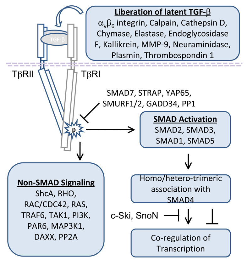
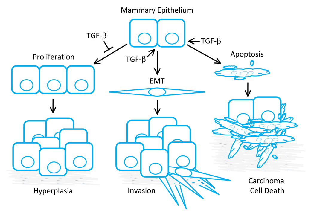
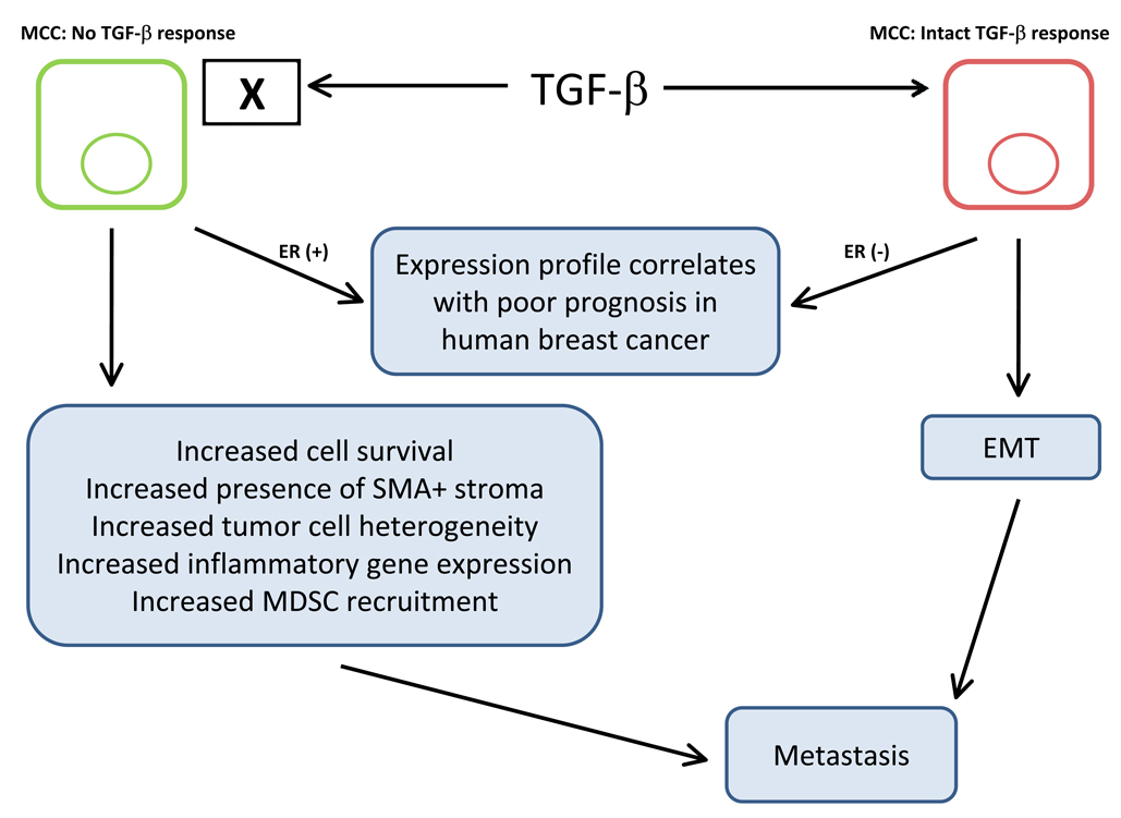
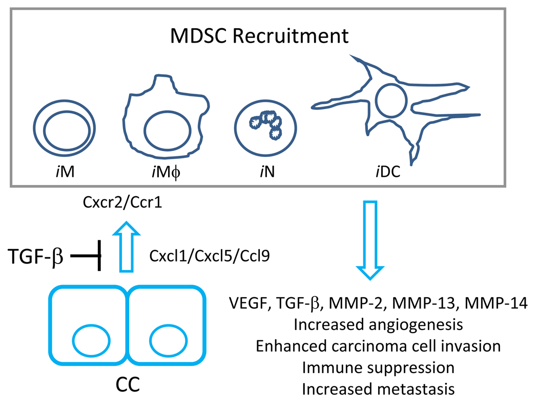
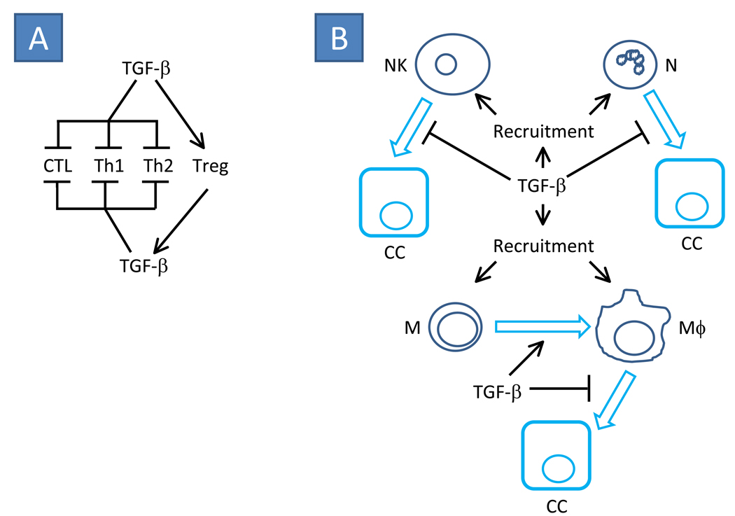
The transforming growth factor beta (TGF-beta) has been studied with regard to the regulation of cell behavior for over three decades. A large body of research has been devoted to the regulation of epithelial cell and derivative carcinoma cell populations in vitro and in vivo. TGF-beta has been shown to inhibit epithelial cell cycle progression and promote apoptosis that together significantly contribute to the tumor suppressive role for TGF-beta during carcinoma initiation and progression. TGF-beta is also able to promote an epithelial to mesenchymal transition that has been associated with increased tumor cell motility, invasion and metastasis. However, it has now been shown that loss of carcinoma cell responsiveness to TGF-beta stimulation can also promote metastasis. Interestingly, enhanced metastasis in the absence of a carcinoma cell response to TGF-beta stimulation has been shown to involve increased chemokine production resulting in recruitment of pro-metastatic myeloid derived suppressor cell (MDSC) populations to the tumor microenvironment at the leading invasive edge. When present, MDSCs enhance angiogenesis, promote immune tolerance and provide matrix degrading enzymes that promote tumor progression and metastasis. Further, the recruitment of MDSC populations in this context likely enhances the classic role for TGF-beta in immune suppression since the MDSCs are an abundant source of TGF-beta production. Importantly, it is now clear that carcinoma-immune cell cross-talk initiated by TGF-beta signaling within the carcinoma cell is a significant determinant worth consideration when designing therapeutic strategies to manage tumor progression and metastasis.


Figures

Figure 1
Figure 1. General overview of TGF-β signaling
TGF-β can be liberated from latent complexes by αvβ6 integrin, calpain, cathepsin D, chymase, elastase, endoglycosidase F, kallikrein, matrix metalloproteinase 9 (MMP-9), neuraminidase, plasmin or thrombospondin 1. Once activated TGF-β is able to bind type II TGF-β receptor homo-dimers (TβRII) thereby permitting the efficient transactivation of type I TGF-β receptor homo-dimers (TβRI). As a result of receptor activation by TGF-β, downstream SMAD dependent and SMAD independent signaling is initiated. However, the level of downstream pathway activation is dependent the abundance of receptor associated TGF-β signaling repressors such as SMAD7, STRAP, YAP65, SMURF1, SMURF2, GADD34 and PP1. SMAD dependent signaling has been primarily associated with activation of SMAD2 or SMAD3, however it has now been shown that SMAD1 and SMAD5 can be activated by TGF-β. Once SMAD1, SMAD2, SMAD3 or SMAD5 are activated they are able to associate with SMAD4 to co-activate or co-repress transcription. Importantly, SMAD dependent signaling can also be repressed by complex association with other transcription factors, co-activators or co-repressors such the c-Ski or SnoN proto-oncogenes. The SMAD independent pathways are known to include ShcA, RHO, RAC/CDC42, RAS, TRAF6, TAK1, PI3K, PAR6, MAP3K1, DAXX and PP2A. Together, the balance of SMAD dependent signaling, SMAD independent signaling and the presence or absence of signaling repressors ultimately determines the response to TGF-β in vitro or in vivo.
Figure 2
Figure 2. Classic roles for TGF-β in the regulation of mammary epithelial cell growth and tumorigenesis
TGF-β signaling has been shown to regulate many epithelial cell types, however many of the common features of signaling through this pathway have been demonstrated using mammary epithelium. TGF-β has been shown to inhibit epithelial cell proliferation predominantly through Smad dependent regulation of p107 (Rb) activity via regulation of p15INK4B, p16INK4A, p21Cip1 and p27Kip1 expression. In the presence of TGF-β, these cyclin dependent kinase inhibitors are upregulated and Rb remains hypo-phosphorylated thereby blocking cell cycle progression. TGF-β is also known to suppress c-myc expression that would otherwise provide an additional proliferative signal for the epithelium. However, one or more of these mediators are often mis-regulated during tumor progression and this can prevent the cytostatic regulation associated with TGF-β stimulation. In addition to cell cycle regulation, TGF-β is able to promote an epithelial to mesenchymal transition (EMT) that has been associated with increased motility and enhanced carcinoma cell invasion. Further, epithelial and carcinoma cell survival has also been associated with TGF-β signaling. Apoptosis induced by TGF-β can provide an additional level of regulation in favor of tumor suppression. The precise role for TGF-β in each epithelial cell population depends on the specific cell type and context of stimulation. Although non-transformed cells from a given organ under clearly defined conditions will often respond to TGF-β in a similar manner, due to the complex nature of parallel intrinsic and extrinsic signaling unique to the cancer evolution in each patient, it is often difficult to predict the proliferative, EMT, or apoptotic impact of TGF-β signaling in a carcinoma cell population a priori.
Figure 3
Figure 3. Gain or loss of TGF-β signaling in mammary carcinoma cells (MCC) can promote metastasis
Gene expression profiles representing gain or loss of TGF-β signaling in mammary carcinoma cells have been shown to correlate with poor prognosis in human breast cancer. Importantly, the results suggest that the difference may be correlated with the status of estrogen receptor (ER) expression. In the presence of ER, a gene signature representing complete abrogation of TGF-β signaling in mammary carcinoma cells correlates with poor clinical prognosis whereas a signature associated with intact TGF-β signaling in ER negative breast cancer correlates with poor clinical prognosis. In the case of intact TGF-β signaling, many studies have suggested that TGF-β dependent EMT promotes carcinoma invasion that initiates and promotes the seeding of distant metastases. In the absence of carcinoma cell specific TGF-β responsiveness, a number of interactions including increased carcinoma cell survival, the increased abundance of adjacent smooth muscle actin positive (SMA+) fibrovascular stroma, increased tumor cell heterogeneity, increased inflammatory gene expression and increased MDSC recruitment have been suggested to promote metastatic progression. Importantly, in a mixed population of carcinoma cells that differ in the ability to respond to TGF-β, it is possible that both types of pro-metastatic progression can co-exist in a single tumor microenvironment.
Figure 4
Figure 4. TGF-β signaling significantly regulates recruitment of tumor promoting myeloid derived suppressor cell (MDSC) populations
MDSC recruitment significantly enhances tumor progression and metastasis. This cell population is a heterogenous combination of myeloid derived cell populations that have characteristics associated with immature monocytes (iM), macrophages (iMϕ), neutrophils (iN) and dendritic cells (iDC). TGF-β signaling in non-transformed and carcinoma-associated epithelial cells (CC) has been shown to suppress the production of chemokines including Cxcl1, Cxcl5 and Ccl9 that when present have the ability to recruit MDSC populations. Cxcl1 and Cxcl5 signal through the Cxcr2 receptor and Ccl9 is known to signal through Ccr1. Abrogation of TGF-β signaling in carcinoma cells relieve the repression of these chemokines and thereby promotes Cxcr2 and Ccr1 dependent recruitment of MDSCs. The recruited MDSC populations are known to be an abundant source of VEGF, TGF-β, MMP-2, MMP-13 and MMP-14 that likely contribute to the MDSC-associated increase in angiogenesis, carcinoma cell invasion, immune suppression and metastasis.
Figure 5
Figure 5. Classic roles for TGF-β signaling in immune suppression often associated with carcinoma-associated tumor progression
(A) TGF-β is known to suppress the activity of cytotoxic T-lymphocytes (CTL) in addition to differentiation of helper Th1 and Th2 T-cell populations. However, TGF-β promotes the differentiation of regulatory T-cells (Treg) that are known to be an abundant source of TGF-β production that may further support suppression of CTL, Th1 and Th2 cells while promoting Treg expansion. (B) TGF-β is a potent chemoattractant for natural killer (NK), neutrophil (N), monocyte (M) and macrophage (Mϕ) cells. TGF-β has been shown to enhance M to Mϕ differentiation. However when present, TGF-β has been shown to prevent NK-, N- and Mϕ-associated carcinoma cell (CC) death. Together, the recruitment of immune cell populations and suppression of immune effector function can result in a tumor microenvironment that is rich in immune cell derived growth factors, morphogens, mitogens, and additional chemokines that further promote tumor progression and metastasis.

Similar articles

Cited by

References

    1. Kehrl JH, et al. Production of transforming growth factor beta by human T lymphocytes and its potential role in the regulation of T cell growth. J Exp Med. 1986;163(5):1037–1050. - PMC - PubMed
    1. Kehrl JH, et al. Transforming growth factor beta is an important immunomodulatory protein for human B lymphocytes. J Immunol. 1986;137(12):3855–3860. - PubMed
    1. Rook AH, et al. Effects of transforming growth factor beta on the functions of natural killer cells: depressed cytolytic activity and blunting of interferon responsiveness. J Immunol. 1986;136(10):3916–3920. - PubMed
    1. Fontana A, et al. Expression of TGF-beta 2 in human glioblastoma: a role in resistance to immune rejection? Ciba Found Symp. 1991;157:232–238. discussion 238-41. - PubMed
    1. Mule JJ, et al. Transforming growth factor-beta inhibits the in vitro generation of lymphokine-activated killer cells and cytotoxic T cells. Cancer Immunol Immunother. 1988;26(2):95–100. - PMC - PubMed

Substances