. 2010 Feb;152(2):553-65.
doi: 10.1104/pp.109.148643. Epub 2009 Dec 16.

Systematic localization of the Arabidopsis core cell cycle proteins reveals novel cell division complexes

Affiliations

Systematic localization of the Arabidopsis core cell cycle proteins reveals novel cell division complexes

Joanna Boruc et al. Plant Physiol. 2010 Feb.

Abstract

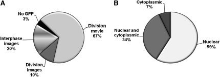
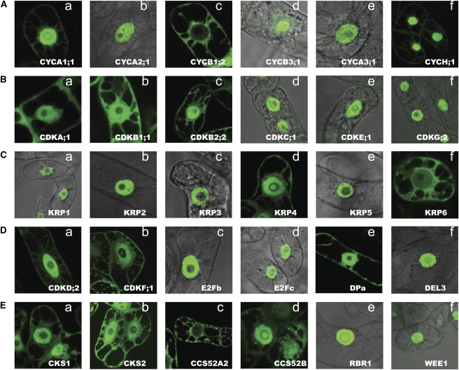
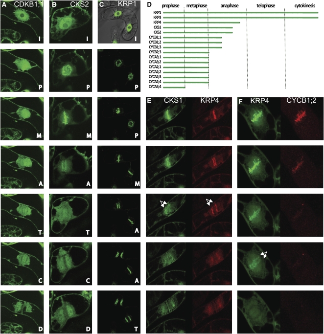
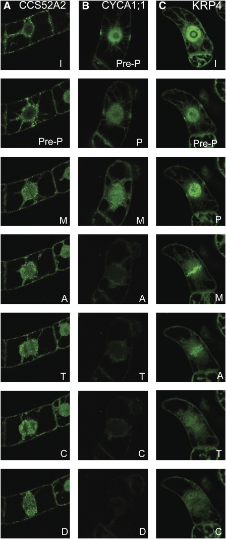
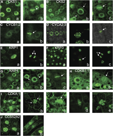
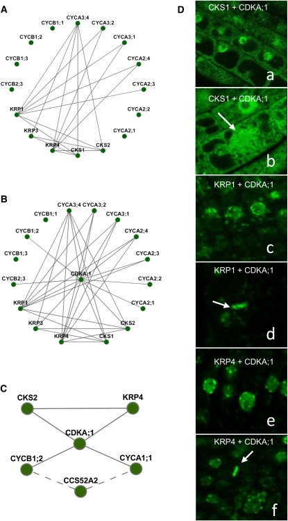
Cell division depends on the correct localization of the cyclin-dependent kinases that are regulated by phosphorylation, cyclin proteolysis, and protein-protein interactions. Although immunological assays can define cell cycle protein abundance and localization, they are not suitable for detecting the dynamic rearrangements of molecular components during cell division. Here, we applied an in vivo approach to trace the subcellular localization of 60 Arabidopsis (Arabidopsis thaliana) core cell cycle proteins fused to green fluorescent proteins during cell division in tobacco (Nicotiana tabacum) and Arabidopsis. Several cell cycle proteins showed a dynamic association with mitotic structures, such as condensed chromosomes and the preprophase band in both species, suggesting a strong conservation of targeting mechanisms. Furthermore, colocalized proteins were shown to bind in vivo, strengthening their localization-function connection. Thus, we identified unknown spatiotemporal territories where functional cell cycle protein interactions are most likely to occur.


Figures

Figure 1.
Figure 1.
The localization patterns of the Arabidopsis cell cycle proteins in interphase cells. A, Pie chart representing 60 analyzed proteins according to their imaging status. B, Interphase localization distribution of analyzed cell cycle proteins.
Figure 2.
Figure 2.
Interphase subcellular localization of selected GFP-tagged proteins representing patterns of localization in BY2. Subcellular localization is shown for cyclin proteins (A), CDKs (B), KRPs (C), CDK-activating kinases and transcription factors (D), and CKS proteins and two CCS52s, RBR1 and WEE1 (E).
Figure 3.
Figure 3.
A to C, Time-lapse analysis of subcellular localization in BY2 cells: CDKB1;1 (A), CKS2 (B), and KRP1 (C). Labels are as follows from top: I, interphase; P, prophase; M, metaphase; A, anaphase; T, telophase; C, cytokinesis; D, daughter cells after division. D, Schematic representation of the timing of protein association with chromosomes and disappearance of the GFP signal from these structures. E and F, Colocalization analysis of chromosomally associated proteins in BY cells: CKS1-GFP and KRP4-RFP (E) and KRP4-GFP and cyclin B1;2-RFP (F). Arrows indicate chromosomes in anaphase.
Figure 4.
Figure 4.
Subcellular localization of proteins associating with the PPB: CCS52A2 (A), CYCA1;1 (B), and KRP4 (C). Labels are as follows from top: I, interphase; Pre-P, preprophase; P, prophase; M, metaphase; A, anaphase; T, telophase; C, cytokinesis; D, daughter cells after division.
Figure 5.
Figure 5.
Subcellular localization of GFP-tagged cell cycle proteins driven by the 35S promoter in Arabidopsis plants. A, CKS1. B, CKS2. C, CYCB1;2. D, CYCA2;3. E, KRP1. F, KRP3. G, KRP4. H, CDKB1;1. I, CDKA;1. J, CCS52A2. Solid arrows indicate metaphase and anaphase chromosomes, and dashed arrows point to the preprophase band signal.
Figure 6.
Figure 6.
Binary protein-protein interaction analysis by the BiFC assay. A and B, Interaction networks of chromosomally associated proteins (A) and the PPI network of chromosomally associated proteins with CDKA;1 (B). C, Interaction network of PPB-associated proteins. Solid lines indicate the interactions detected in this study with the BiFC assay, and dashed lines depict previously published interactions (with the coimmunoprecipitation assay). D, Confocal images of Arabidopsis cells coexpressing binding protein complexes tested in the BiFC assay. Subcellular localization of complexes is as follows: CKS1 and CDKA;1 (a and b), KRP1 and CDKA;1 (c and d), KRP4 and CDKA;1 (e and f), and KRP3 and CDKA;1 (g). Arrows indicate cells in metaphase.

References

    1. Bähler J (2005) Cell-cycle control of gene expression in budding and fission yeast. Annu Rev Genet 39 69–94 - PubMed
    1. Bird DA, Buruiana MM, Zhou Y, Fowke LC, Wang H (2007) Arabidopsis cyclin-dependent kinase inhibitors are nuclear-localized and show different localization patterns within the nucleoplasm. Plant Cell Rep 26 861–872 - PubMed
    1. Boudolf V, Lammens T, Boruc J, Van Leene J, Van Den Daele H, Maes S, Van Isterdael G, Russinova E, Kondorosi E, Witters E, et al (2009) CDKB1;1 forms a functional complex with CYCA2;3 to suppress endocycle onset. Plant Physiol 150 1482–1493 - PMC - PubMed
    1. Bourne Y, Watson MH, Hickey MJ, Holmes W, Rocque W, Reed SI, Tainer JA (1996) Crystal structure and mutational analysis of the human CDK2 kinase complex with cell cycle-regulatory protein CksHs1. Cell 84 863–874 - PubMed
    1. Breyne P, Zabeau M (2001) Genome-wide expression analysis of plant cell cycle modulated genes. Curr Opin Plant Biol 4 136–142 - PubMed

Publication types

MeSH terms

LinkOut - more resources