. 2010 Jan 15;184(2):702-12.
doi: 10.4049/jimmunol.0902360. Epub 2009 Dec 16.

Gene expression analysis of macrophages that facilitate tumor invasion supports a role for Wnt-signaling in mediating their activity in primary mammary tumors

Affiliations

Gene expression analysis of macrophages that facilitate tumor invasion supports a role for Wnt-signaling in mediating their activity in primary mammary tumors

Laureen S Ojalvo et al. J Immunol. .

Abstract

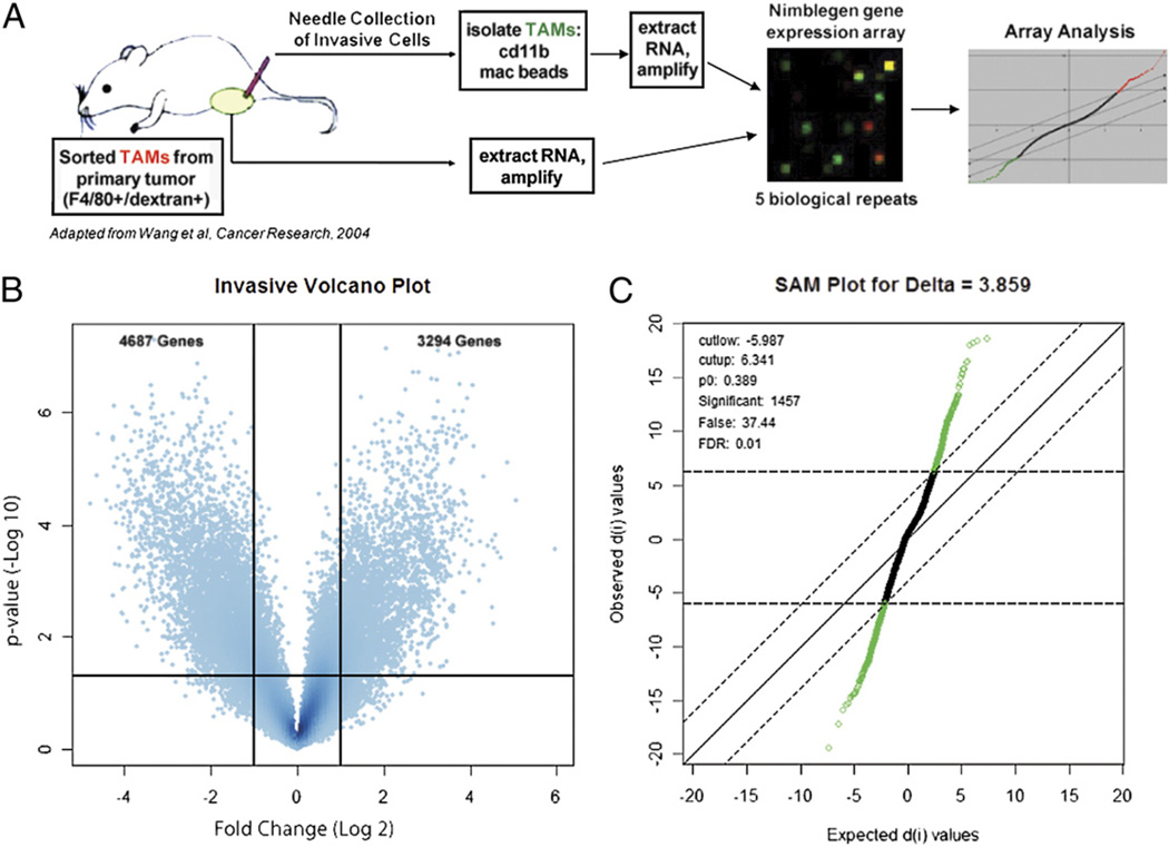
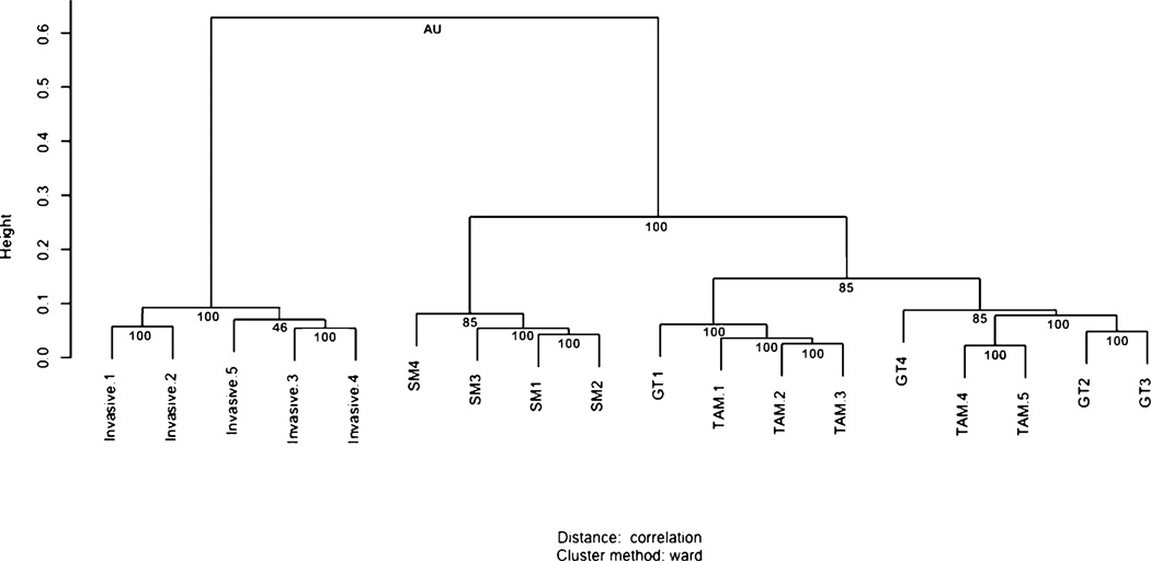
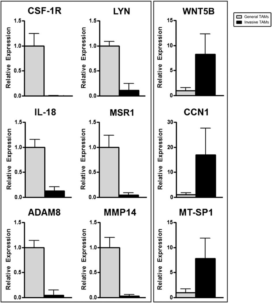
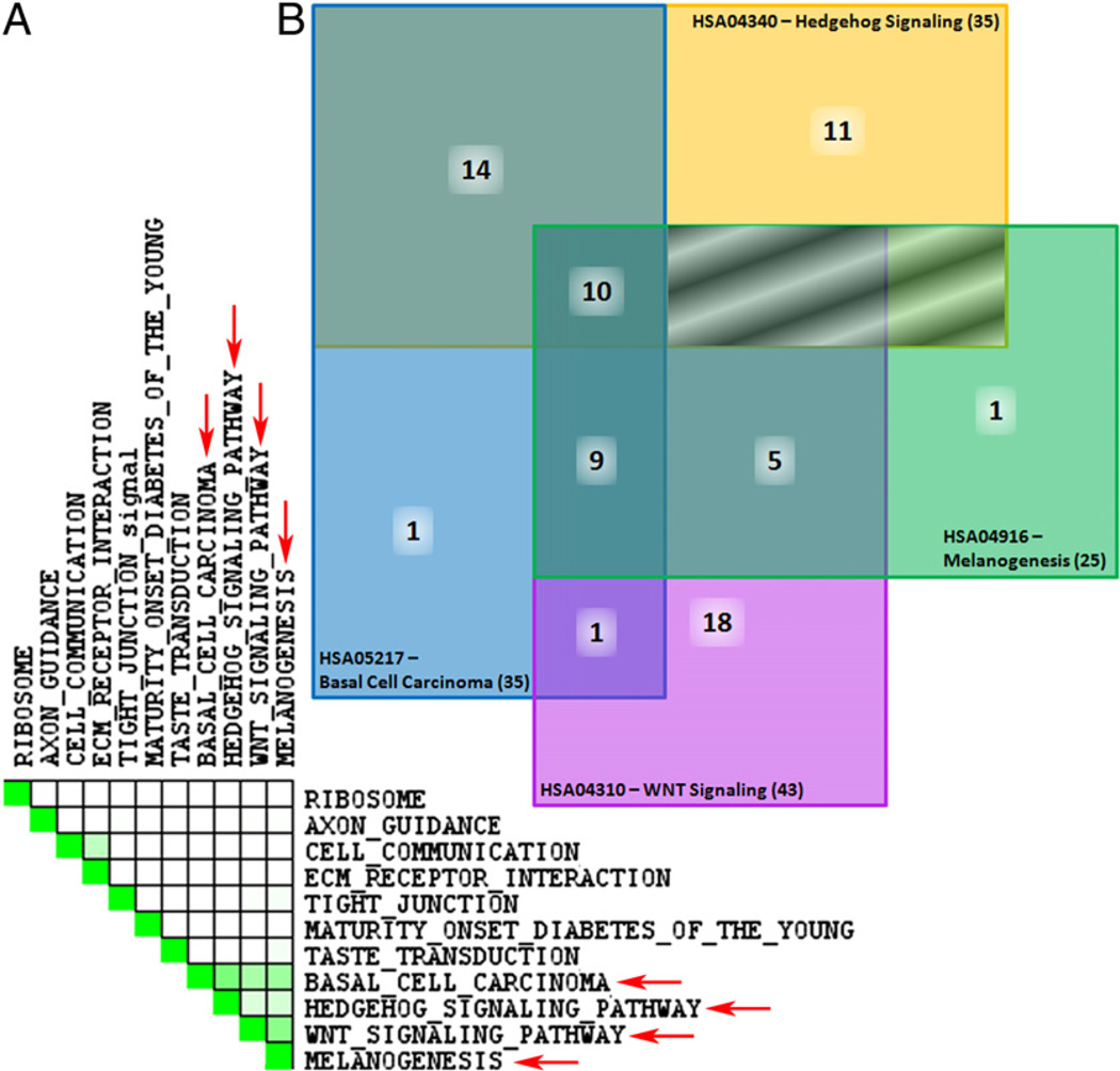
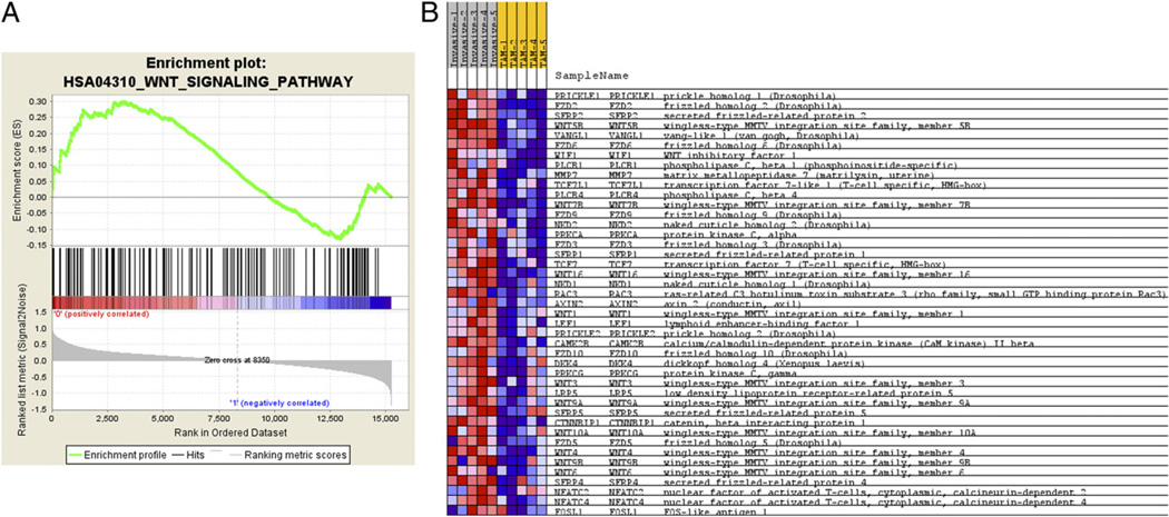
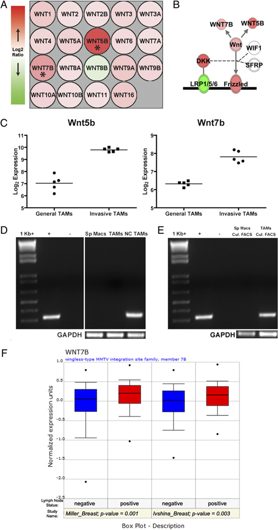
The tumor microenvironment modifies the malignancy of tumors. In solid tumors, this environment is populated by many macrophages that, in genetic studies that depleted these cells from mouse models of breast cancer, were shown to promote tumor progression to malignancy and increase metastatic potential. Mechanistic studies showed that these tumor-promoting effects of macrophages are through the stimulation of tumor cell migration, invasion, intravasation, and enhancement of angiogenesis. Using an in vivo invasion assay, it was demonstrated that invasive carcinoma cells are a unique subpopulation of tumor cells whose invasion and chemotaxis is dependent on the comigration of tumor-associated macrophages (TAMs) with obligate reciprocal signaling through an epidermal growth factor-CSF-1 paracrine loop. In this study, these invasion-promoting macrophages were isolated and subjected to analysis of their transcriptome in comparison with TAMs isolated indiscriminately to function using established macrophage markers. Unsupervised analysis of transcript patterns showed that the invasion-associated TAMs represent a unique subpopulation of TAMs that, by gene ontology criteria, have gene expression patterns related to tissue and organ development. Gene set enrichment analysis showed that these macrophages are also specifically enriched for molecules involved in Wnt-signaling. Previously, it was shown that macrophage-derived Wnt molecules promote vascular remodeling and that tumor cells are highly motile and intravasate around perivascular TAM clusters. Taken together, we conjecture that invasive TAMs link angiogenesis and tumor invasion and that Wnt-signaling plays a role in mediating their activity.


Conflict of interest statement

Disclosures

The authors have no financial conflicts of interest.

Figures

FIGURE 1
FIGURE 1
Isolation of a functionally defined subpopulation of invasive TAMs for gene expression analysis. A, Schematic of array experimental protocol comparing gene expression profile of invasive TAMs to GTs (26). B, Volcano plot illustrating fold change (log base 2) compared with p value (−log base 10) between invasive TAMs and GTs. Horizontal bar at y ≈ 1.303 represents a significance level of p = 0.05 significance level. C, SAM for δ 3.859, FDR 1%.
FIGURE 2
FIGURE 2
Invasive TAMs are a unique subpopulation of TAMs Unsupervised clustering of data from several macrophage populations shows that invasive TAMs group together in a clade with strong statistical support. A total of 18 macrophage samples were used including the five invasive TAM samples (Invasive) and five GT samples from the FVB mouse strain used in this study, and the four TAM and 4 splenic macrophage (SM) samples from mice on a mixed background, as previously published (4). Approximately unbiased (AU) scores are listed below individual pairs. Values close to 100 indicate a reliable clade. Larger heights on the vertical axis are indicative of a greater distance between clades.
FIGURE 3
FIGURE 3
QRT-PCR validates gene expression array data for downregulated (A) and upregulated (B) transcripts. Invasive TAMs (black bars) and GTs (gray bars) normalized to expression of housekeeping gene cyclophilin A (Ppia).
FIGURE 4
FIGURE 4
KEGG pathways enriched in invasive TAMs are related through Wnts. A, Set-to-set diagram, created through performing a leading edge analysis using GSEA, indicates the overlap between significantly enriched KEGG pathways listed in Table II. Dark green cells indicate that the gene sets have leading edge genes in common, whereas a white cell indicates no leading edge gene overlap. Four sets demonstrate overlap (red arrows). B, Venn diagram of overlapping molecules between enriched KEGG pathways. Notably, 10 molecules are common to all four pathways including: Wnts 1, 3, 4, 5B, 6, 7B, 9A, 9B, 10A and 16. Boxes shaded using diagonal lines have no overlap between pathways. Official human KEGG pathway signifiers are listed.
FIGURE 5
FIGURE 5
GSEA analysis for Wnt-signaling pathway and positively enriched associated molecules. A, Distribution of the 142 Wnt-signaling pathway molecules amid the total ranked list of all transcripts analyzed by GSEA (46). Enrichment score of the pathway of 0.299 is identified by determining the peak of positively correlated transcripts. B, Normogram for the core enriched Wnt-signaling pathway related molecules in the array dataset. Increased expression (red), decreased expression (blue).
FIGURE 6
FIGURE 6
Wnt7b in TAMs and human breast cancer. A, Expression pattern for all Wnt ligands queried by Nimblegen gene expression arrays. Most Wnt ligands were found to be increased (red) in invasive TAMs compared with general TAMs, except for Wnt8b which was decreased (green). Asterisks indicate Wnt5b and Wnt7b that were called as significantly differentially regulated. B, Generalized representation of relative abundance of molecules driving Wnt-signaling in invasive TAMs. Double outlined molecules (Frizzled, LRP1/5/6, DKK, and WNT) contain multiple members that are not individually depicted. Shading represents the trend for the entire group (e.g., note expression of individual Wnt ligands in A versus total WNT expression). Diagram generated using IPA 6.3. C, Normalized array expression intensities (log base 2) for Wnt5b and Wnt7b for each biologic replicate depict reproducibility between samples. The line indicates average expression for each set of experiments.D, Semiquantitative PCR for Wnt7b expression in splenic macrophages (Sp Macs), GTs (TAMs) and invasive, needle-collected TAMs (NC TAMs). E, Representative semiquantitative PCR for Wnt7b in splenic macrophages (Sp Macs) and TAMs following flow cytometric sorting and 12-h culture to ensure pure macrophage populations. F, Higher expression of Wnt7b in human breast cancer correlates with advanced disease. Two separate studies were mined using Oncomine (39) and indicate that breast cancer metastatis to lymph nodes has significantly higher WNT7B expression.

References

    1. Pollard JW. Trophic macrophages in development and disease. Nat. Rev. Immunol. 2009;9:259–270. - PMC - PubMed
    1. Joyce JA, Pollard JW. Microenvironmental regulation of metastasis. Nat. Rev. Cancer. 2009;9:239–252. - PMC - PubMed
    1. Pollard JW. Tumour-educated macrophages promote tumour progression and metastasis. Nat. Rev. Cancer. 2004;4:71–78. - PubMed
    1. Ojalvo LS, King W, Cox D, Pollard JW. High-density gene expression analysis of tumor-associated macrophages from mouse mammary tumors. Am. J. Pathol. 2009;174:1048–1064. - PMC - PubMed
    1. Lewis CE, Pollard JW. Distinct role of macrophages in different tumor microenvironments. Cancer Res. 2006;66:605–612. - PubMed

Publication types