. 2010 Jan 5;107(1):314-9.
doi: 10.1073/pnas.0910547107. Epub 2009 Dec 14.

Ebolavirus VP35 uses a bimodal strategy to bind dsRNA for innate immune suppression

Affiliations

Ebolavirus VP35 uses a bimodal strategy to bind dsRNA for innate immune suppression

Christopher R Kimberlin et al. Proc Natl Acad Sci U S A. .

Abstract

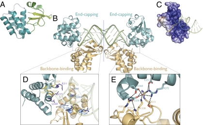
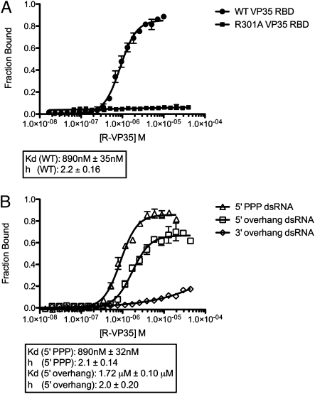
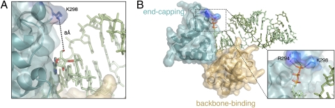
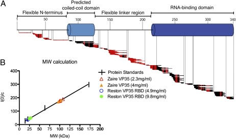
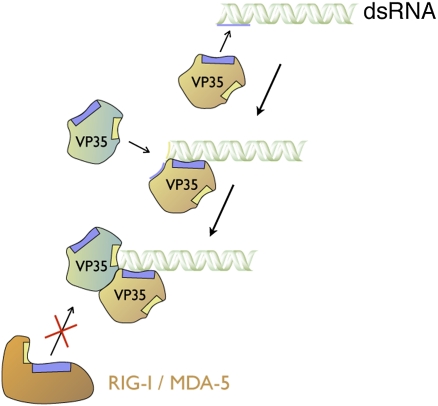
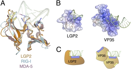
Ebolavirus causes a severe hemorrhagic fever and is divided into five distinct species, of which Reston ebolavirus is uniquely nonpathogenic to humans. Disease caused by ebolavirus is marked by early immunosuppression of innate immune signaling events, involving silencing and sequestration of double-stranded RNA (dsRNA) by the viral protein VP35. Here we present unbound and dsRNA-bound crystal structures of the dsRNA-binding domain of Reston ebolavirus VP35. The structures show that VP35 forms an unusual, asymmetric dimer on dsRNA binding, with each of the monomers binding dsRNA in a different way: one binds the backbone whereas the other caps the terminus. Additional SAXS, DXMS, and dsRNA-binding experiments presented here support a model of cooperative dsRNA recognition in which binding of the first monomer assists binding of the next monomer of the oligomeric VP35 protein. This work illustrates how ebolavirus VP35 could mask key recognition sites of molecules such as RIG-I, MDA-5, and Dicer to silence viral dsRNA in infection.


Conflict of interest statement

The authors declare no conflict of interest.

Figures

Fig. 1.
Fig. 1.
(A) Subdomain organization of the unbound VP35 RBD monomer. The α-helical subdomain is teal and the β-sheet subdomain is green. (B) Asymmetric unit of the VP35 RBD-dsRNA complex structure. Two VP35 RBDs (backbone-binding, orange; end-capping, teal) bind as an asymmetric dimer to each end of an 18-bp dsRNA oligomer (green). A pseudo 2-fold axis runs through the midpoint of the dsRNA. (C) Electrostatic surface potential of the asymmetric VP35 RBD dimer (colored continuously) highlighting the basic pocket into which blunt-ended dsRNA binds. Yellow line denotes the boundary between VP35 monomers. Positive surface is colored blue; negative surface is colored red, with limits ±10 kT/e. (D) Both the backbone-binding RBD (orange) and end-capping RBD (teal) contain polar residues (backbone-binding, blue; end-capping, purple) that interact with the dsRNA phosphate backbone. Only the end-capping RBD also uses hydrophobic residues (yellow) to form a nonpolar face that packs against the terminal bases of the dsRNA. (E) Polar residues at the dimeric interface between the backbone-binding (orange) and end-capping (teal) RBDs with possible hydrogen bonds (dashed black lines).
Fig. 2.
Fig. 2.
RNA binding studies of VP35 RBD. (A) Dual-filter dot blot experiments with blunt-ended 18bp dsRNA indicate that the wild-type Reston VP35 RBD (filled circles, n = 60) binds cooperatively, with a Hill coefficient of 2.2 ± 0.16 SEM. By contrast, the R301A point mutant of the Reston VP35 RBD (filled squares, n = 40) abrogates dsRNA binding. (B) Wild-type Reston VP35 RBD binds dsRNA with 5′ triphosphate (open triangles, n = 40) and 5′ overhangs (open squares, n = 60) with affinity and cooperativity similar to that of blunt-ended dsRNA. The VP35 RBD retains limited ability to bind dsRNA with 3′ overhangs (open diamonds, n = 60), but affinity is greatly reduced.
Fig. 3.
Fig. 3.
Triphosphate modeled onto the 5′ end of the dsRNA in the dsRNA-dimeric VP35 RBD complex. (A) Although the dsRNA crystallized contains 5′ hydroxyls, adequate space exists to accommodate a 5′ triphosphate and VP35 indeed binds 5′ triphosphate-bearing dsRNA. (B) VP35-dsRNA complex with a triphosphate modeled onto the 5′ terminus of the dsRNA. VP35 residues R294 and K298 appear to be well positioned to hydrogen bond with a 5′ triphosphate.
Fig. 4.
Fig. 4.
Biophysical characterization of VP35. (A) Ten-second amide hydrogen–deuterium exchange map for select VP35 peptides of a near full-length Zaire VP35. Red horizontal bars indicate fast-exchanging amides; black bars indicate stretches of no exchange. (B) Molecular weight calculation of Zaire VP35 (triangles) and the Reston VP35 RBD (circles). Our Zaire VP35 expression construct encodes a 37-k Da molecule. SAXS analysis indicates a ∼100 kDa mass, consistent with the oligomeric (probably trimeric) nature of VP35 in solution. The RBD construct (here, indicated by Reston) encodes a molecule ∼20 kDa in size. SAXS indicates a ∼20–25 kDa mass, consistent with the monomeric nature of the RBD in the absence of dsRNA.
Fig. 5.
Fig. 5.
Similar modes of binding between host dsRNA receptors and VP35 RBD. (A) Superimposition of the RIG-I C-terminal domain (CTD) (pale cyan; PDB 2QFB) and MDA-5 CTD (mauve; PDB 3GA3) with the LGP2 CTD (brown; PDB 3EQT), highlighting the structural similarity among the three homologs. Conserved polar residues (blue) make key contacts with the phosphate backbone in the LGP2-dsRNA complex structure, whereas hydrophobic residues (yellow) pack against terminal nucleotide bases of the dsRNA (transparent green). (B) Electrostatic surface potential of the LGP2 CTD (colored continuously) bound to an 8-bp dsRNA oligo (green) highlighting surface charge distribution similar to that of the asymmetric VP35 dimer. Positive surface is colored blue; negative surface is colored red with limits ± 10 kT/e. (C) Schematic view of LGP2 CTD and VP35 RBD dimer, illustrating organization of polar (blue) and hydrophobic (yellow) interaction sites for dsRNA.
Fig. 6.
Fig. 6.
Model of the cooperative, dimeric assembly mechanism of the VP35 RBD on dsRNA. The interface between the dsRNA and the backbone-binding VP35 RBD is larger (540 Å2) than either the dsRNA-end-capping RBD interface (340 Å2), or the RBD-RBD interface in the dimer (330 Å2). Furthermore, the affinity for uncapped and 5′triphosphate-capped dsRNA is similar, suggesting that a 5′ triphosphate is not a major recognition signal. These results and the observed in-solution cooperativity of dsRNA binding together suggest a mechanism by which the backbone-binding VP35 forms the first and initial contact to the phosphate backbone along the side of the dsRNA (brown VP35, blue box to blue bar). The second VP35 RBD (the end-capping molecule) would then bind the larger surface now assembled by both the terminus of the RNA and the dimer interface of the backbone-binding VP35 (teal VP35, yellow box to dsRNA end). In this model, the assembly of the dimeric VP35 RBDs on the end of the dsRNA oligonucleotide masks the binding site of RIG-I and MDA-5, preventing their recognition of dsRNA and subsequent signaling.

Similar articles

Cited by

References

    1. Sanchez A, Geisbert TW, Feldmann H. Filoviridae: Marburg and Ebola Viruses. In: Knipe DM, Howley PM, editors. Fields Virology. Philidelphia: Lippincott Williams & Wilkins; 2007. pp. 1409–1448.
    1. Towner JS, et al. Newly discovered ebola virus associated with hemorrhagic fever outbreak in Uganda. PLoS Pathog. 2008;4:e1000212. - PMC - PubMed
    1. Bwaka MA, et al. Ebola hemorrhagic fever in Kikwit, Democratic Republic of the Congo: Clinical observations in 103 patients. J Infect Dis. 1999;179(Suppl 1):S1–S7. - PubMed
    1. Barrette RW, et al. Discovery of swine as a host for the Reston ebolavirus. Science. 2009;325:204–206. - PubMed
    1. CDC. Update: Filovirus infections among persons with occupational exposure to nonhuman primates. MMWR Morb Mortal Wkly. 1990;39:266–7. 273. - PubMed

Publication types

MeSH terms

Substances

LinkOut - more resources