. 2010 Jan 5;107(1):64-8.
doi: 10.1073/pnas.0911164107. Epub 2009 Dec 14.

Recombination-induced tag exchange to track old and new proteins

Affiliations

Recombination-induced tag exchange to track old and new proteins

Kitty F Verzijlbergen et al. Proc Natl Acad Sci U S A. .

Abstract

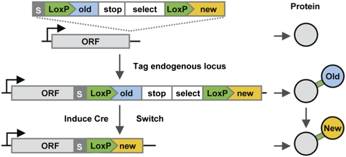
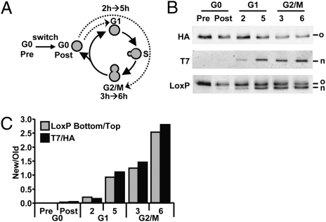
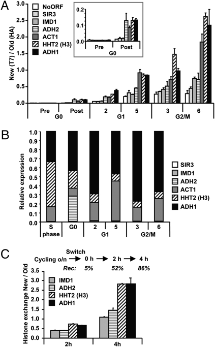
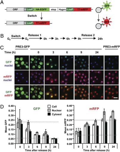
The dynamic behavior of proteins is critical for cellular homeostasis. However, analyzing dynamics of proteins and protein complexes in vivo has been difficult. Here we describe recombination-induced tag exchange (RITE), a genetic method that induces a permanent epitope-tag switch in the coding sequence after a hormone-induced activation of Cre recombinase. The time-controlled tag switch provides a unique ability to detect and separate old and new proteins in time and space, which opens up opportunities to investigate the dynamic behavior of proteins. We validated the technology by determining exchange of endogenous histones in chromatin by biochemical methods and by visualizing and quantifying replacement of old by new proteasomes in single cells by microscopy. RITE is widely applicable and allows probing spatiotemporal changes in protein properties by multiple methods.


Conflict of interest statement

The authors declare no conflict of interest.

Figures

Fig. 1.
Fig. 1.
Outline of recombination-induced tag exchange (RITE). RITE cassettes contain two epitope tags (old and new), the first of which is in between two LoxP sites. Integration of a RITE cassette downstream of an ORF (ORF) results in a protein tagged with an old tag (blue). The old tag is preceded by an invariant flexible spacer (S) and a short peptide encoded by the LoxP sequence (LoxP) and is followed by a transcriptional terminator (stop) and a selectable marker (select). Upon induction of Cre recombinase, site-specific recombination between the tandem LoxP sites in the genome results in loss of the old tag and fusion of the ORF to the new tag. After the switch, newly synthesized proteins will contain the new tag (yellow), whereas existing proteins will contain the old tag. Old and new proteins are expressed from the same gene by the native promoter.
Fig. 2.
Fig. 2.
Application of RITE to endogenous histone H3. (A) One of the two genes encoding histone H3 in yeast (HHT2) was tagged with a RITE cassette (H3-RITE) containing short epitope tags: HA (old) and T7 (new). The other gene encoding histone H3 (HHT1) was deleted. A Hygromycin resistance gene (Hygro) was used to select against illegitimate recombinants. The tag switch was under control of a constitutively expressed hormone-dependent Cre recombinase (Cre-EBD78). (B) Growth of wild-type and H3 RITE-tagged [before (HA) and after (T7) the switch] yeast cells spotted in a 10-fold dilution series. (C) The efficiency of recombination in the cell population was determined by Southern blot analysis of genomic DNA digested with HindIII (H) before (Pre) and after (Post) addition of the hormone β-estradiol. An invariant fragment was used as a control (Ctrl). (D) Detection of old (HA) and new (T7) histone H3 by quantitative immunoblot analysis of whole cell lysates of equal numbers of starved switched cells released into fresh media. The number of population doublings was calculated by staining the cells with N-hydroxysuccinimide-tetra-ethylrhodamine (NHS-TER) (SI Materials and Methods). (E) The percentage of old H3-HA plotted against the number of population doublings. The measured HA/T7 ratios of the blot in D were converted into H3-HA percentages by using standard curves of samples with known percentages of H3-HA and H3-T7 (SI Materials and Methods).
Fig. 3.
Fig. 3.
Global histone exchange determined by immunodetection. (A) Yeast strains were grown to saturation (here referred to as G0) in complete medium and recombination was induced overnight (switch) by addition of hormone (Fig. S1). Cells were released in fresh media and arrested in G1 (α-factor) or G2/M (nocodazole). Samples were taken at the estimated start of the arrest (2 h G1 and 3 h G2/M) and 3 h later. (B) Quantitative immunoblot analysis of old and new histone H3 in whole-cell lysates using antibodies against HA (old), T7 (new) or an antibody raised against the spacer-LoxP sequence (LoxP) recognizing old and new proteins simultaneously. (C) Relative H3-T7/H3-HA ratios (New/Old) were calculated on the basis of the ratio of the top band (H3-HA) and the bottom band (H3-T7) of the LoxP blot (absolute values) and the ratio of HA and T7 signals (arbitrary units).
Fig. 4.
Fig. 4.
Replication-independent transcription-coupled histone turnover quantified by affinity purification. (A) Analysis of chromatin-bound histones by ChIP of HA (old) and T7 (new) histone H3 quantified by real-time quantitative PCR (qPCR). Histone exchange (ratio of new/old) was determined for promoters of the indicated genes and an intergenic region on chromosome V (NoORF). The genes are ranked by estimated transcription frequency in log phase, from low (open bars) to high (solid bars) frequency. The result shown is the average of two individual experiments (± SEM). The Inset is a zoom-in of the G0 time points. (B) Relative mRNA expression levels were determined by reverse-transcriptase qPCR (RT-qPCR). An S-phase sample of the same H3-RITE strain was used as a reference sample. A wild-type strain without a RITE tag showed very similar expression profiles (Fig. S4). (C) Histone turnover in H3-T7→HA cells without any arrest was determined by induction of Cre-recombinase in log-phase cells (OD660 = 0.25). The percentage of cells that had undergone recombination (Rec) is indicated for each time point (determined by a colony-plating assay). Histone replacement at promoters was determined by ChIP (HA/T7).
Fig. 5.
Fig. 5.
Spatiotemporal analysis of old and new proteasomes by microscopy. (A) Schematic representation of fluorescent RITE. (B) PRE3-GFP→mRFP cells were grown to saturation (G0) and recombination was induced overnight (switch). Subsequently, cells were released in fresh media (release 1) and samples were taken at the indicated time points. Nine hours after the first release, cells were again supplemented with fresh media (release 2). Time points 3, 6, 9, and 24 h correspond to ≈0.3, 2, 3, and 8 cell divisions, respectively. (C) Representative confocal microscopy images of PRE3-GFP→mRFP grown as indicated in B and of control strains (PRE3-GFP and PRE3-mRFP). Hoechst was used as a nuclear counterstaining (blue). (Scale bar, 4 μm.) (D) The GFP and mRFP fluorescent intensities of micrographs from C were quantified and the value shown for each time point is an average of the mean fluorescence intensity in the nuclei, cytoplasm, and total surface of 400 cells (± SD). Dashed lines indicate GFP and mRFP signals in control cells expressing GFP or mRFP only (the bottom dashed lines indicate background levels).

Similar articles

Cited by

References

    1. Gorski SA, Dundr M, Misteli T. The road much traveled: Trafficking in the cell nucleus. Curr Opin Cell Biol. 2006;18:284–290. - PubMed
    1. Russel D, et al. The structural dynamics of macromolecular processes. Curr Opin Cell Biol. 2009;21:97–108. - PMC - PubMed
    1. Reits EA, Neefjes JJ. From fixed to FRAP: Measuring protein mobility and activity in living cells. Nat Cell Biol. 2001;3:E145–E147. - PubMed
    1. D’Angelo MA, Hetzer MW. Structure, dynamics and function of nuclear pore complexes. Trends Cell Biol. 2008;18:456–466. - PMC - PubMed
    1. Adams SR, Tsien RY. Preparation of the membrane-permeant biarsenicals FlAsH-EDT2 and ReAsH-EDT2 for fluorescent labeling of tetracysteine-tagged proteins. Nat Protoc. 2008;3:1527–1534. - PMC - PubMed

Publication types

MeSH terms

LinkOut - more resources