. 2010 Jan 5;107(1):246-51.
doi: 10.1073/pnas.0908428107. Epub 2009 Dec 14.

Mutant p53 drives metastasis and overcomes growth arrest/senescence in pancreatic cancer

Affiliations

Mutant p53 drives metastasis and overcomes growth arrest/senescence in pancreatic cancer

Jennifer P Morton et al. Proc Natl Acad Sci U S A. .

Erratum in

Abstract

TP53 mutation occurs in 50-75% of human pancreatic ductal adenocarcinomas (PDAC) following an initiating activating mutation in the KRAS gene. These p53 mutations frequently result in expression of a stable protein, p53(R175H), rather than complete loss of protein expression. In this study we elucidate the functions of mutant p53 (Trp53(R172H)), compared to knockout p53 (Trp53(fl)), in a mouse model of PDAC. First we find that although Kras(G12D) is one of the major oncogenic drivers of PDAC, most Kras(G12D)-expressing pancreatic cells are selectively lost from the tissue, and those that remain form premalignant lesions. Loss, or mutation, of Trp53 allows retention of the Kras(G12D)-expressing cells and drives rapid progression of these premalignant lesions to PDAC. This progression is consistent with failed growth arrest and/or senescence of premalignant lesions, since a mutant of p53, p53(R172P), which can still induce p21 and cell cycle arrest, is resistant to PDAC formation. Second, we find that despite similar kinetics of primary tumor formation, mutant p53(R172H), as compared with genetic loss of p53, specifically promotes metastasis. Moreover, only mutant p53(R172H)-expressing tumor cells exhibit invasive activity in an in vitro assay. Importantly, in human PDAC, p53 accumulation significantly correlates with lymph node metastasis. In summary, by using 'knock-in' mutations of Trp53 we have identified two critical acquired functions of a stably expressed mutant form of p53 that drive PDAC; first, an escape from Kras(G12D)-induced senescence/growth arrest and second, the promotion of metastasis.


Conflict of interest statement

The authors declare no conflict of interest.

Figures
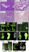
Fig. 1.
Fig. 1.
Noninvasive in vivo imaging in a murine model of pancreatic ductal adenocarcinoma (PDAC). (A–C) H&E staining of (A) a normal pancreatic duct in a Pdx1-Cre-GFP, LSL-Trp53R172H (PC) mouse, (B) a PanIN lesion in a Pdx1-Cre-GFP-LSL-KrasG12D, LSL-Trp53R172H (KPC) mouse, and (C) an invasive adenocarcinoma from a KPC mouse. (D) Alcian blue staining of a PanIN lesion from a KPC mouse. (E and F) In vivo imaging, using the Olympus OV100 in vivo imaging system, of GFP fluorescence within the pancreata of (E) 2-week-old and (F) 3-week-old Pdx1-Cre-GFP, LSL-KrasG12D/+ (KC) mice and PC mice, as indicated. (Top) Whole-body imaging; (Bottom) ×8 magnification of excised pancreata. (G and H) In vivo imaging, using the Olympus OV100 in vivo imaging system, of GFP fluorescence within the pancreata of (G) 4-week-old and (H) 6-week-old KC mice and PC mice, as indicated.
Fig. 2.
Fig. 2.
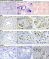
Senescence program is activated in KrasG12D-expressing cells in the normal pancreas but not in pancreatic tumors. (A–C) β-Galactosidase staining at pH 6, (D–F) p53 immunohistochemical staining, (G–I) p21 immunohistochemical staining, and (J–L) MCM2 immunohistochemical staining, in sections of frozen (β-galactosidase) or formalin-fixed paraffin-embedded pancreatic tissue. (A, D, G, and J) Normal pancreatic ducts in pancreata harvested from 6-week-old Pdx1-Cre-GFP, LSL-KrasG12D/+ (KC) mice, (B, E, H, and K) PanIN lesions in pancreata harvested from 6-week-old KC mice, and (C, F, I, and L) pancreatic ductal adenocarcinoma harvested from Pdx1-Cre-GFP-LSL-KrasG12D, LSL-Trp53R172H (KPC) mice. Arrowheads indicate areas where p21 up-regulation has been lost.
Fig. 3.
Fig. 3.
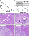
p53 Drives metastasis of pancreatic ductal adenocarcinoma. (A) Kaplan-Meier survival curve shows no significant difference in survival between Pdx1-Cre-GFP, LSL-KrasG12D, LSL-Trp53R172H/+ (KPC) mice (solid line), and Pdx1-Cre-GFP, LSL-KrasG12D/+, LSL-Trp53loxP/+ (KPflC) mice (dashed line). P = 0.479. (B) Table showing that mean lifespan and median survival are not significantly different in KPC mice compared with KPflC mice, whereas incidence of metastasis in KPC mice is significantly increased compared with KPflC mice, in which metastasis is not observed at all. (C) p53 Histoscore in relation to lymph node status in cases of human PDAC (0, lymph node negative; 1, metastatic disease present in <50% of lymph nodes sampled; 2, metastatic disease present in >50% of lymph nodes sampled; mean number of nodes reviewed per resection, 21). (D and E) H&E-stained sections from (D) a KPC tumor and (E) an age-matched KPflC tumor show there is no difference in tumor stage or grade between the two genotypes. (F and G) H&E-stained sections of liver metastases arising in KPC mice.
Fig. 4.
Fig. 4.
Mutant p53 promotes invasion of PDAC cells in vitro. Inverted invasion assays were performed on murine PDAC tumor cell lines with or without mutant p53. (A) Tumor cell lines bearing mutant p53R172H, from Pdx1-Cre-GFP, LSL-KrasG12D, LSL-Trp53R172H/+ (KPC) tumors (Middle), invade further than tumor cells grown from Pdx1-Cre-GFP, LSL-KrasG12D/+, LSL-Trp53loxP/+ (KPflC) tumors (Top). Introduction of exogenous expression of mutant p53R175H into these KPflC tumor cells, however, promotes invasion (Bottom). (B) Bar graph showing increased invasive capacity of KPC tumor cells compared with KPflC tumor cells. Representative images of at least three independent experiments are shown. Columns indicate mean; bars indicate SE. *P ≤ 0.01. (C) Western immunoblotting shows expression of flag-tagged p53R175H in stably transfected KPflC cells. β-Tubulin represents a loading control. (D) Bar graph showing increased invasive capacity of KPflC tumor cells following exogenous expression of mutant p53R175H. Representative images of at least three independent experiments are shown. Columns indicate mean; bars indicate SE. *P < 0.01 by unpaired Student’s t test.

References

    1. Warshaw AL, Fernández-del Castillo C. Pancreatic carcinoma. N Engl J Med. 1992;326:455–465. - PubMed
    1. Jensen OM, Estève J, Møller H, Renard H. Cancer in the European Community and its member states. Eur J Cancer. 1990;26:1167–1256. - PubMed
    1. Hruban RH, Goggins M, Parsons J, Kern SE. Progression model for pancreatic cancer. Clin Cancer Res. 2000;6:2969–2972. - PubMed
    1. Hingorani SR, et al. Trp53R172H and KrasG12D cooperate to promote chromosomal instability and widely metastatic pancreatic ductal adenocarcinoma in mice. Cancer Cell. 2005;7:469–483. - PubMed
    1. Almoguera C, et al. Most human carcinomas of the exocrine pancreas contain mutant c-K-ras genes. Cell. 1988;53:549–554. - PubMed

Publication types

MeSH terms

Substances