. 2009 Dec 29;106(52):22329-34.
doi: 10.1073/pnas.0907464106. Epub 2009 Dec 11.

Planarian Hedgehog/Patched establishes anterior-posterior polarity by regulating Wnt signaling

Affiliations

Planarian Hedgehog/Patched establishes anterior-posterior polarity by regulating Wnt signaling

Shigenobu Yazawa et al. Proc Natl Acad Sci U S A. .

Abstract

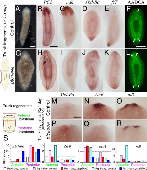
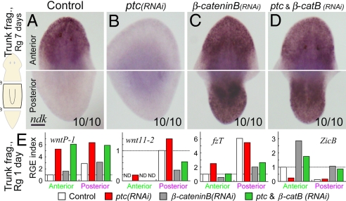
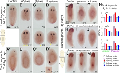
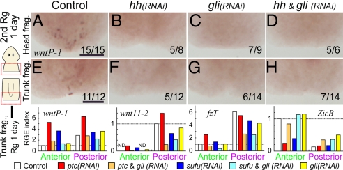
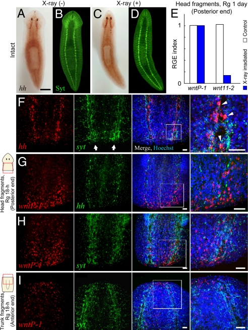
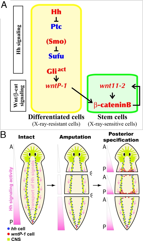
Despite long-standing interest, the molecular mechanisms underlying the establishment of anterior-posterior (AP) polarity remain among the unsolved mysteries in metazoans. In the planarians (a family of flatworms), canonical Wnt/beta-catenin signaling is required for posterior specification, as it is in many animals. However, the molecular mechanisms regulating the posterior-specific induction of Wnt genes according to the AP polarity have remained unclear. Here, we demonstrate that Hedgehog (Hh) signaling is responsible for the establishment of AP polarity via its regulation of the transcription of Wnt family genes during planarian regeneration. We found that RNAi gene knockdown of Dugesia japonica patched (Djptc) caused ectopic tail formation in the anterior blastema of body fragments, resulting in bipolar-tails regeneration. In contrast, RNAi of hedgehog (Djhh) and gli (Djgli) caused bipolar-heads regeneration. We show that Patched-mediated Hh signaling was crucial for posterior specification, which is established by regulating the transcription of Wnt genes via downstream Gli activity. Moreover, differentiated cells were responsible for the posterior specification of undifferentiated stem cells through Wnt/beta-catenin signaling. Surprisingly, Djhh was expressed in neural cells all along the ventral nerve cords (along the AP axis), but not in the posterior blastema of body fragments, where the expression of Wnt genes was induced for posteriorization. We therefore propose that Hh signals direct head or tail regeneration according to the AP polarity, which is established by Hh signaling activity along the body's preexisting nervous system.


Conflict of interest statement

The authors declare no conflict of interest.

Figures

Fig. 1.
Fig. 1.
“Janus-tails” phenotype is caused by Djptc RNAi. Anterior end is placed at the top in all panels. Rg, regeneration. (A–L) Djptc(RNAi) caused abnormal anterior blastema formation in trunk fragment regenerants at 7–9 days postamputation. (A and G) Live animals. (B–F and H–L) Characterization by in situ detection of DjPC2 (CNS marker; B and H), Djndk (a head marker; C and I), DjAbd-Ba (a tail marker; D and J), and DjfzT (a tail marker; E and K), and by immunostaining for DjAADCA (digestive gut branch staining; F and L). Arrowheads indicate the single anterior and two posterior gut branches (F), and the bifurcated branching of the main gut tract in the anterior region as posterior fate (L). Arrow indicates an extra pharynx (H). (Scale bars, 500 μm.) (M–R) Posteriorized anterior end in Djptc(RNAi) trunk regenerants 1 day after amputation. Expression patterns of DjAbd-Ba (M and P), DjZicB (anterior marker; N and Q), and Djndk (O and R) in the anterior ends. (Scale bar, 200 μm.) (S) Time course of relative gene expression (RGE) level reveals gradual posteriorization of anterior blastema after Djptc(RNAi) during early regeneration (1 and 3 days). The expression levels of target genes (DjAbd-Ba, DjZicB, DjotxA, and Djndk) in anterior or posterior blastemas were normalized and expressed relative to the level in the anterior end of control trunk regenerants at day 0 taken as 1.0 (white column of “Anterior” bars; indicated by dashed line). Mean of quadruplicates.
Fig. 2.
Fig. 2.
Djptc functions upstream of Wnt/β-catenin signaling. (A–D) Djndk expression in anterior (Upper) and posterior (Lower) regions of RNAi trunk regenerants at day 7. Djptc(RNAi) caused Janus-tails formation (B), whereas Djβ-cateninB(RNAi) caused Janus-heads formation (C) with or without simultaneous Djptc(RNAi) (D). Numbers represent the fraction of tested regenerants showing the indicated phenotype. (Scale bar, 200 μm.) (E) RGE levels of Wnt genes in anterior and posterior blastemas of RNAi trunk regenerants at day 1, with the anterior (DjZicB) and posterior (DjfzT) marker genes shown as positive controls for RNAi phenotype. ND, not detected.
Fig. 3.
Fig. 3.
Hh signaling is involved in the establishment of AP polarity. (A–D) Loss of Hh signaling activity caused Janus-heads or loss of the tail identity in regeneration. DjfzT expression in head fragments at day 11 of the first round of regeneration (A–D), and trunk fragments at day 7 of the second round of regeneration (A′–D′) after strong inhibition of the positive regulators of the Hh signaling cascade (RNAi of Djhh or Djgli). Note that the generation of ectopic eyes in the posterior blastemas (arrowheads) and extra eye formation (arrow; Inset) near the posterior end (7%; 1/14, D′). (Scale bars, 300 μm.) (E–M) Janus-tails formation after RNAi of negative regulators of Hh signaling cascade (Djptc or Djsufu) and its suppression by simultaneous RNAi of positive regulators. Djndk expression in trunk fragments at regeneration day 9. (Scale bar, 300 μm.) (N) RGE index of Hh signaling component genes in anterior and posterior blastemas after Djptc(RNAi) as in Fig. 1S.
Fig. 4.
Fig. 4.
Hh signaling regulates transcription of Wnt genes. (A–H) DjwntP-1 expression was lost by RNAi of Djhh or Djgli in the posterior end of both head (A–D) and trunk (E–H) regenerants on day 1 of the second round of regeneration. (Scale bars, 200 μm.) (I) RGE quantification of Wnt genes in anterior and posterior blastemas of trunk fragments at regeneration day 1 after RNAi of Hh signaling components genes, with the anterior (DjZicB) and posterior (DjfzT) marker genes as positive controls for RNAi phenotype. ND, not detected.
Fig. 5.
Fig. 5.
Posteriorizing signal originates in differentiated cells (neuron). (A–D) Expression of Djhh was observed in X-ray-resistant differentiated cells of the VNCs. In situ detection of Djhh in nonirradiated (A) and irradiated (C) planarians, followed by labeling of the CNS with anti-DjSynaptotagimin antibody (neural marker; Syt; B and D). (Scale bar, 1 mm.) (E) RGE index of Wnt genes in the posterior blastema of head fragments from X-ray-irradiated animals 1 day after amputation. (F-I) Confocal images of expression patterns of Djhh, DjwntP-1, or Djsyt by dual in situ detection in the posterior (F–H) and anterior (I) end in regenerants from head and trunk fragments, respectively, at 18 h after amputation. Panels show individual expression patterns (red or green color) and their merged image with the image of nuclear staining (Hoechst; blue). The rightmost panel is a magnified view of each boxed area. Arrow indicates VNC (F). Note that Djhh was expressed in neural cells of the VNCs, which were labeled with Djsyt probe (arrowheads; F), but not in the posterior blastema (F and G). Also, DjwntP-1 was predominantly expressed in the posterior end surrounding the VNCs (G and H), but was also weakly expressed in the anterior end (I). (Scale bars, 50 μm.)
Fig. 6.
Fig. 6.
Models for two aspect of the establishment of AP polarity by Hh signaling in planarians. (A) The signaling cascade model for posterior specification in planarian regeneration inferred from the present study. Hh signaling triggers posteriorization by inducing transcription of Wnt genes in differentiated cells (yellow-shaded box), from which the signal is transmitted to stem cells (green-shaded box) through Wnt/β-cateninB signaling for tail regeneration. Positive and negative regulators of posterior specification are written with red and blue letters, respectively. (B) A possible model for the establishment of AP polarity by Hh signaling in planarians. DjwntP-1-expressing cells (red dots) at the posterior end suggests that Hh signaling is exclusively activated there (pink gradients on the left), although Djhh-expressing cells (neural cells; blue dots) are localized all along the VNCs (CNS is indicated by light green). In this model, Djhh gene products would be transported posteriorly via VNCs (pink arrows), leading to the induction of posterior specification at the posterior end.

References

    1. Morgan TH. An analysis of the phenomena of organic ‘polarity’. Science. 1904;20:742–748. - PubMed
    1. Morgan TH. The control of heteromorphosis in Planaria maculata. Arch Entw Mech Org. 1904;17:683–695.
    1. Reddien PW, Sánchez Alvarado A. Fundamentals of planarian regeneration. Annu Rev Cell Dev Biol. 2004;20:725–757. - PubMed
    1. Gurley KA, Rink JC, Sánchez Alvarado A. β-catenin defines head versus tail identity during planarian regeneration and homeostasis. Science. 2008;319:323–327. - PMC - PubMed
    1. McWhinnie MA. The effect of colchicine on reconstitutional development in Dugesia dorotocephala. Bio Bull. 1955;108:54–65.

Publication types

MeSH terms