. 2009 Dec 29;106(52):22381-6.
doi: 10.1073/pnas.0906372106. Epub 2009 Dec 16.

Mast cells regulate homeostatic intestinal epithelial migration and barrier function by a chymase/Mcpt4-dependent mechanism

Affiliations

Mast cells regulate homeostatic intestinal epithelial migration and barrier function by a chymase/Mcpt4-dependent mechanism

Katherine R Groschwitz et al. Proc Natl Acad Sci U S A. .

Abstract

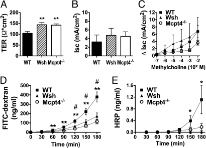
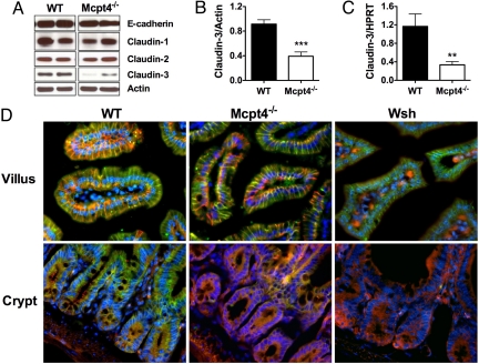
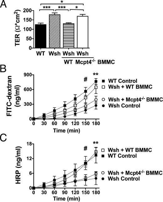
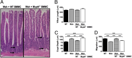
Altered intestinal barrier function is postulated to be a central predisposing factor to intestinal diseases, including inflammatory bowel diseases and food allergies. However, the mechanisms involved in maintaining homeostatic intestinal barrier integrity remain undefined. In this study, we demonstrate that mice deficient in mast cells (Kit(W-sh/W-sh) [Wsh]) or mast cell chymase (Mcpt4(-/-)) have significantly decreased basal small intestinal permeability compared with wild-type (WT) mice. Altered intestinal barrier function was linked to decreased intestinal epithelial cell migration along the villus/crypt axis, altered intestinal morphology, and dysregulated claudin-3 crypt expression. Remarkably, engraftment of Wsh mice with WT but not Mcpt4(-/-) mast cells restored intestinal epithelial cell migration, morphology, and intestinal epithelial barrier function. Collectively, these findings identify a mechanism by which mast cells regulate homeostatic intestinal epithelial migration and barrier function.


Conflict of interest statement

The authors declare no conflict of interest.

Figures

Fig. 1.
Fig. 1.
Decreased basal intestinal permeability in Wsh and Mcpt4−/− mice. Segments of jejunum from WT, Wsh, and Mcpt4−/− mice were mounted in Ussing chambers and the baseline (A) transepithelial resistance (TER) and (B) short-circuit current (Isc), and (C) β-methylcholine-stimulated changes in Isc were measured. Ex vivo intestinal permeability was measured as luminal-to-serosal flux of (D) FITC-dextran and (E) HRP. Values represent mean± SEM; n = 12–18 mice per group. Statistical significance is: (A) **, P < 0.01 vs. WT; (D) #, P < 0.01 Wsh vs. WT, **, P < 0.01 Mcpt4−/− vs. WT; (E) *, P < 0.05 Wsh vs. WT.
Fig. 2.
Fig. 2.
Altered intestinal architecture and epithelial migration in Wsh and Mcpt4−/− mice. (A) H&E stained jejunum from WT, Wsh, and Mcpt4−/− mice was examined for (B) villus length, measured from villus-crypt junction to villus tip and (C) crypt depth, measured from crypt base to villus-crypt junction. (A) Arrows demonstrate approximate crypt measurements and highlight the difference between mice. (D) Epithelial migration was measured as the distance (μm) from crypt base to the farthest migrated BRDU+ cell. Values represent mean± SEM; n = 5–7 mice per group. Statistical significance is: (C) *, P < 0.01 and ***, P < 0.001 vs. WT; (D) **, P < 0.01 vs. WT.
Fig. 3.
Fig. 3.
Decreased claudin-3 expression in jejunal crypt epithelium of Mcpt4−/− and Wsh mice. (A) E-cadherin, claudin-1, -2, and -3 and actin were evaluated by Western blot. (B) Densitometric analysis of claudin-3 expression normalized to β-actin. (C) Quantitative real-time PCR analysis of claudin-3 normalized to HPRT. (D) Claudin-3 (green) and E-cadherin (red) immunofluorescence of the villi (Top) and crypts (Bottom) of jejunum from WT (Left), Mcpt4−/− (Middle), and Wsh (Right) mice; nuclei stained by DAPI (blue). Western representative of three individual experiments. Densitometry represents mean ± SEM; n = 10 mice. PCR represents n = 6–8 mice per group. Statistical significance is: **, P < 0.01, ***, P < 0.001.
Fig. 4.
Fig. 4.
Mast cell engraftment of Wsh mice with WT, but not Mcpt4−/− BMMCs restores basal intestinal permeability. Wsh mice were engrafted with BMMCs from WT or Mcpt4−/− mice. Twelve weeks later, jejunum was examined ex vivo for (A) TER and luminal-to-serosal flux of (B) FITC-dextran and (C) HRP. Values represent mean± SEM; n = 4–8 mice per group. Representative of three independent experiments. Statistical significance by two-way ANOVA is: (A) *, P < 0.05, ***, P < 0.001; (B) #, P < 0.01 WT control vs. Wsh control; **, P < 0.001 WT control vs. Wsh control, P < 0.05 WT control vs. Mcpt4−/− BMMC and P < 0.01 Wsh control vs. WT BMMC; (C) #, P < 0.05 WT control vs. Wsh control, WT control vs. KO BMMC, WT BMMC vs. Wsh control and WT BMMC vs. KO BMMC; **, P < 0.001 WT control vs. Wsh control, WT BMMC vs. Wsh control and WT BMMC vs. KO BMMC, and P < 0.01 WT control vs. KO BMMC.
Fig. 5.
Fig. 5.
Mast cell engraftment of Wsh mice with WT, but not Mcpt4−/− BMMCs restores intestinal architecture and epithelial migration. Wsh mice were engrafted with BMMCs from WT or Mcpt4−/− mice and examined after 12 weeks. H&E-stained jejunum from (A) Wsh mice engrafted with either WT or Mcpt4−/− BMMCs were examined for (B) villus length, measured from villus-crypt junction to villus tip and (C) crypt depth, measured from crypt base to villus-crypt junction. Arrows demonstrate approximate location of crypt measurements and highlight the difference between mice. (D) Mice were injected with 0.2 mg/g body weight BRDU and after 24 h, epithelial migration was measured as the distance from the crypt base to the farthest migrated BRDU+ cell. Values represent mean± SEM; n = 4–8 mice per group. Statistical significance is: **, P < 0.01, ***, P < 0.001.

Similar articles

Cited by

References

    1. Berkes J, Viswanathan VK, Savkovic SD, Hecht G. Intestinal epithelial responses to enteric pathogens: Effects on the tight junction barrier, ion transport, and inflammation. Gut. 2003;52:439–451. - PMC - PubMed
    1. Groschwitz KR, Hogan SP. Intestinal barrier function: Molecular regulation and disease pathogenesis. J Allergy Clin Immunol. 2009;124:3–20. - PMC - PubMed
    1. Peeters M, et al. Clustering of increased small intestinal permeability in families with Crohn's disease. Gastroenterology. 1997;113:802–807. - PubMed
    1. Soderholm JD, et al. Different intestinal permeability patterns in relatives and spouses of patients with Crohn's disease: An inherited defect in mucosal defence? Gut. 1999;44:96–100. - PMC - PubMed
    1. van Elburg RM, Uil JJ, Mulder CJ, Heymans HS. Intestinal permeability in patients with celiac disease and relatives of patients with celiac disease. Gut. 1993;34:354–357. - PMC - PubMed

Publication types

MeSH terms