. 2010 Jan 5;107(1):75-80.
doi: 10.1073/pnas.0912589107. Epub 2009 Dec 15.

Isolation and characterization of centroacinar/terminal ductal progenitor cells in adult mouse pancreas

Affiliations

Isolation and characterization of centroacinar/terminal ductal progenitor cells in adult mouse pancreas

Meritxell Rovira et al. Proc Natl Acad Sci U S A. .

Abstract

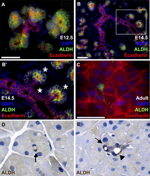
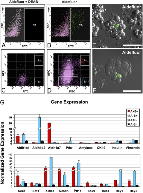
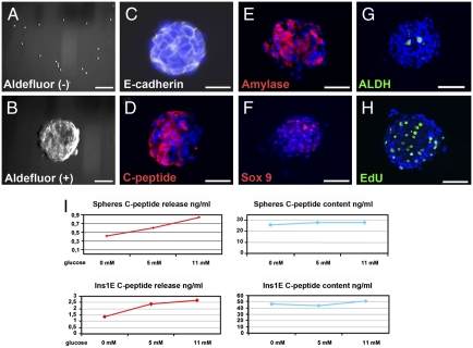
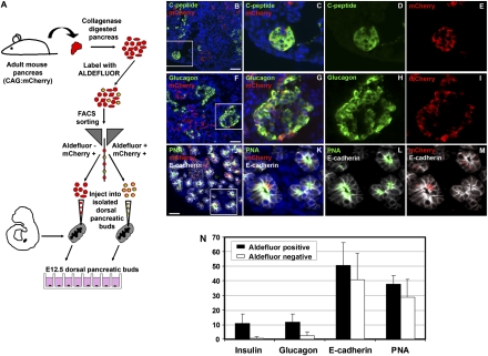
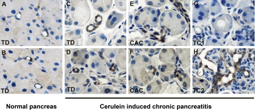
The question of whether dedicated progenitor cells exist in adult vertebrate pancreas remains controversial. Centroacinar cells and terminal duct (CA/TD) cells lie at the junction between peripheral acinar cells and the adjacent ductal epithelium, and are frequently included among cell types proposed as candidate pancreatic progenitors. However these cells have not previously been isolated in a manner that allows formal assessment of their progenitor capacities. We have found that a subset of adult CA/TD cells are characterized by high levels of ALDH1 enzymatic activity, related to high-level expression of both Aldh1a1 and Aldh1a7. This allows their isolation by FACS using a fluorogenic ALDH1 substrate. FACS-isolated CA/TD cells are relatively depleted of transcripts associated with differentiated pancreatic cell types. In contrast, they are markedly enriched for transcripts encoding Sca1, Sdf1, c-Met, Nestin, and Sox9, markers previously associated with progenitor populations in embryonic pancreas and other tissues. FACS-sorted CA/TD cells are uniquely able to form self-renewing "pancreatospheres" in suspension culture, even when plated at clonal density. These spheres display a capacity for spontaneous endocrine and exocrine differentiation, as well as glucose-responsive insulin secretion. In addition, when injected into cultured embryonic dorsal pancreatic buds, these adult cells display a unique capacity to contribute to both the embryonic endocrine and exocrine lineages. Finally, these cells demonstrate dramatic expansion in the setting of chronic epithelial injury. These findings suggest that CA/TD cells are indeed capable of progenitor function and may contribute to the maintenance of tissue homeostasis in adult mouse pancreas.


Conflict of interest statement

The authors declare no conflict of interest.

Figures

Fig. 1.
Fig. 1.
ALDH1 expression in embryonic and adult mouse pancreas. (A–C) Immunofluorescent labeling for ALDH1 protein (green) in combination with E-cadherin (red) to mark epithelial structures in E12.5 (A), E14.5 (B and B′), and adult mouse pancreas (C). Image in (B′) represents higher magnification view of area indicated by box in (B). Note restriction of ALDH1 expression to tips of epithelial branches (indicated by asterisks in B′) and not more-central branch trunks (indicated by star). In adult pancreas (C), ALDH1 expression is restricted to a subset of E-cadherin-positive centroacinar cells. (D and E) Immunohistochemical detection of ALDH1 protein (brown) in subsets of centroacinar (arrows) and terminal duct cells (arrowhead). (Scale bars: 50 μM.)
Fig. 2.
Fig. 2.
FACS isolation of ALDH1-expressing centroacinar/terminal ductal epithelial cells using the Aldefluor reagent. FACS sorting was performed on single cells isolated from peripheral acinar-ductal units depleted of endocrine and large duct elements. (A and B) Gating of Aldefluor-positive cells based on DEAB-sensitive ALDH1 enzymatic activity. y axis indicates side scatter; x axis indicates intensity of Aldefluor signal (A) with and (B) without DEAB. (B and C) Detection of ALDH1 enzymatic activity (C) with and (D) without DEAB, in conjunction with surface detection of E-cadherin protein. y axis represents intensity of labeling with APC-conjugated anti-E-cadherin antibody; x axis indicates intensity of Aldefluor signal. FACS-sorted populations indicated by P2, P3, P4, and P5 in D correspond to Aldefluor-positive, E-cadherin-negative (A+E−), Aldefluor-positive, E-cadherin-positive (A+E+), Aldefluor-negative, E-cadherin-positive (A−E+), and Aldefluor-negative, E-cadherin-negative (A−E−), respectively. (E and F) Imaging of collagenase-digested mouse pancreas using Aldefluor reagent confirms centroacinar/terminal ductal localization of Aldefluor-positive cells, similar to that observed for ALDH1 immunofluorescence (Figs. 1 and 2). Note centroacinar/terminal ductal position and small size of Aldefluor-positive cells relative to larger acinar cells, which are easily identifiable by granular cytoplasm corresponding to apical zymogen granules. (Scale bars: 50 μM.) (G) Quantitative RT-PCR analysis of gene expression in A+E+ cells (red), A+E− cells (white), A+E− cells (blue), and A−E− cells (black). Compared with A−E+ aldefluor-negative epithelial cells, A+E+ aldefluor-positive centroacinar/terminal ductal epithelial cells are enriched for transcripts encoding Aldh1a1, Aldh1a7, Sca1, Sdf1, c-Met, Nestin, Ptf1a, and Sox9. (Scale bars: 50 μM.)
Fig. 3.
Fig. 3.
Formation, differentiation, and function of pancreatospheres derived from Aldefluor-positive centroacinar/terminal ductal cells. (A and B) A+E+ centroacinar/terminal ductal epithelial cells, but not A−E+ epithelial cells, efficiently form pancreatospheres in suspension culture. (C–G) Expression of E-cadherin (C), insulin C-peptide (D), amylase (E), Sox9 (F), and ALDH1 (G) in day 7 pancreatospheres formed from A+E+ centroacinar/terminal ductal epithelial cells. (H) Cell proliferation in day 7 pancreatospheres as assessed by overnight incorporation of EdU added on day 6 of culture period. (I) ELISA-based assay of stored and secreted insulin C-peptide following overnight incubation of either pancreatospheres or Ins-1 cells in varying concentrations of glucose. Note that pancreatospheres display glucose sensitivity similar to that observed in Ins-1 cells (i.e., ∼2-fold increase in secreted C-peptide in response to 0 vs. 11 mM glucose). (Scale bars: 100 μM.)
Fig. 4.
Fig. 4.
Aldefluor-positive adult pancreatic cells enter both endocrine and exocrine lineages in cultured embryonic pancreas. (A) Schematic of experiment. To trace the lineage of adult cells, Aldefluor (+) and Aldefluor (–) cells were isolated from adult CAG:mCherry transgenic mouse pancreas, microinjected into microdissected dorsal pancreatic buds isolated from E12.5 nontransgenic mouse embryos, and assayed for an ability to productively contribute to the developing endocrine and exocrine lineages. (B–J) Coexpression of mCherry and insulin C-peptide (B–E) and mCherry and glucagon (F–I) confirms capacity of adult Aldefluor (+) cells to contribute to embryonic β- and α-cell lineages, whereas labeling of individual mCherry-positive cells with FITC-conjugated PNA (J–M) confirms ability to contribute to the embryonic acinar lineage. (N) Frequencies with which residual mCherry-positive adult Aldeflouor (+) and Aldefluor (−) cells label for insulin C-peptide, glucagon, E-cadherin, and PNA 7 days after microinjection into microdissected E12.5 dorsal pancreatic buds. All cell counts were determined using E-cadherin labeling to outline the boundary of individual cells. Note that the capacity for endocrine differentiation is predominantly limited to the Aldefluor (+) population, whereas both Aldefluor (+) and Aldefluor (−) cells can productively contribute to the developing exocrine lineages. (Scale bars: 50 μM.)
Fig. 5.
Fig. 5.
Expansion of ALDH1-expressing centroacinar and terminal ductal epithelial cells in setting of chronic inflammation and regenerative epithelial metaplasia. Following antigen retrieval, ALDH1 protein was detected using immunohistochemistry on pancreatic tissue from normal adult pancreas (A and B) and pancreas harvested from mice with chronic pancreatitis induced by three weekly injections of caerulein (C–H). (A and B) Low-frequency labeling for ALDH1 in terminal ductal (TD) epithelial cells from normal adult pancreas. (C and D) Expansion of ALDH1-expressing terminal ductal epithelium following sequential caerulein administration. (E and F) Similar expansion of ALDH1-expressing centroacinar cells (CAC) following sequential caerulein administration. (G and H) Expression of ALDH1 in caerulein-induced metaplastic type 2 (TC2; H), but not type 1 (TC1; G) tubular complexes.

References

    1. Zhou Q, Brown J, Kanarek A, Rajagopal J, Melton DA. vivo reprogramming of adult pancreatic exocrine cells to beta-cells. Nature. 2008;455:627–632. - PMC - PubMed
    1. Sangiorgi E, Capecchi MR. Bmi1 lineage tracing identifies a self-renewing pancreatic acinar cell subpopulation capable of maintaining pancreatic organ homeostasis. Proc Natl Acad Sci USA. 2009;106:7101–7106. - PMC - PubMed
    1. Dor Y, Brown J, Martinez OI, Melton DA. Adult pancreatic beta-cells are formed by self-duplication rather than stem-cell differentiation. Nature. 2004;429:41–46. - PubMed
    1. Fendrich V, et al. Hedgehog signaling is required for effective regeneration of exocrine pancreas. Gastroenterology. 2008;135:621–631. - PMC - PubMed
    1. Teta M, Rankin MM, Long SY, Stein GM, Kushner JA. Growth and regeneration of adult beta cells does not involve specialized progenitors. Dev Cell. 2007;12:817–826. - PubMed

Publication types

MeSH terms