. 2010 Jan 5;107(1):436-41.
doi: 10.1073/pnas.0908099107. Epub 2009 Dec 15.

NCLX is an essential component of mitochondrial Na+/Ca2+ exchange

Affiliations

NCLX is an essential component of mitochondrial Na+/Ca2+ exchange

Raz Palty et al. Proc Natl Acad Sci U S A. .

Abstract

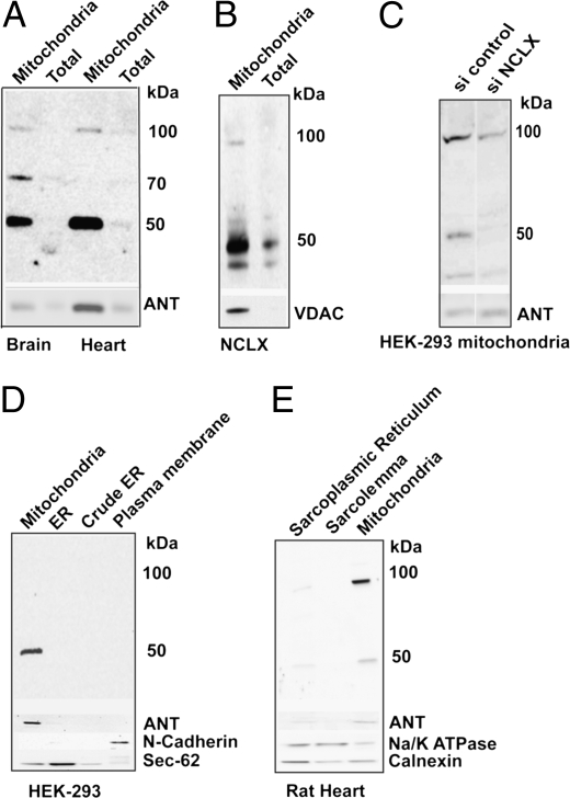
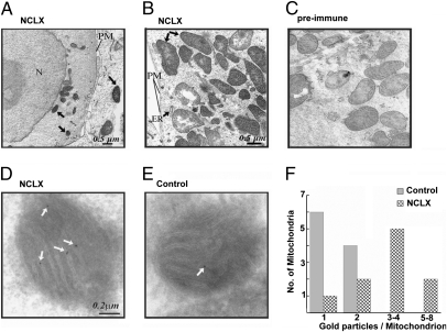
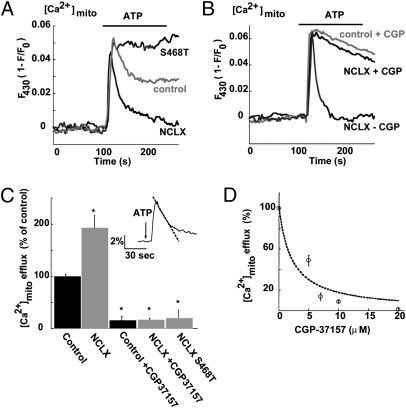
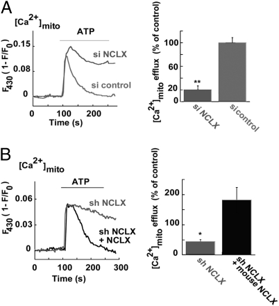
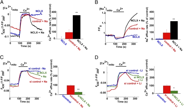
Mitochondrial Ca(2+) efflux is linked to numerous cellular activities and pathophysiological processes. Although it is established that an Na(+)-dependent mechanism mediates mitochondrial Ca(2+) efflux, the molecular identity of this transporter has remained elusive. Here we show that the Na(+)/Ca(2+) exchanger NCLX is enriched in mitochondria, where it is localized to the cristae. Employing Ca(2+) and Na(+) fluorescent imaging, we demonstrate that mitochondrial Na(+)-dependent Ca(2+) efflux is enhanced upon overexpression of NCLX, is reduced by silencing of NCLX expression by siRNA, and is fully rescued by the concomitant expression of heterologous NCLX. NCLX-mediated mitochondrial Ca(2+) transport was inhibited, moreover, by CGP-37157 and exhibited Li(+) dependence, both hallmarks of mitochondrial Na(+)-dependent Ca(2+) efflux. Finally, NCLX-mediated mitochondrial Ca(2+) exchange is blocked in cells expressing a catalytically inactive NCLX mutant. Taken together, our results converge to the conclusion that NCLX is the long-sought mitochondrial Na(+)/Ca(2+) exchanger.


Conflict of interest statement

The authors declare no conflict of interest.

Figures

Fig. 1.
Fig. 1.
NCLX is localized to mitochondria. (A) Immunoblot analysis of NCLX in total tissue lysate (Total) and mitochondrial fractions (Mitochondria) purified from mouse heart and brain (15 μg). (B) Immunoblot analysis of total cellular and mitochondrial fractions purified from HEK-293-T cells overexpressing murine NCLX (10 μg). (Lower) Immunoblots of ANT or VDAC serving as markers. (C) Immunoblot analysis of NCLX in mitochondrial fractions purified from HEK-293 cells transfected with siNCLX or scrambled siRNA (siControl). Note that siNCLX diminishes expression levels of both the 50-kDa and 100-kDa forms of NCLX. (D and E) Expression of NCLX in cellular and tissue fractions of the indicated components purified from HEK-293 cells (20 μg) (D) or rat heart (10 μg) (E). (Lower) Immunoblots of ANT (mitochondrial marker), Na+/K+ ATPase, or N-cadherin (plasma membrane, PM, marker) and Calnexin or Sec-62 (ER, marker). Note that the mitochondria are the major site of NCLX localization (the slight NCLX signal in cardiac sarcoplasmic reticulum is presumably related to cross-contamination with mitochondria; see ANT staining).
Fig. 2.
Fig. 2.
NCLX is found on the inner membrane of mitochondria. (A–C) Electron micrographs of rat cortical slices (A) or CHO cells (B) stained with anti-NCLX antibodies or NCLX preimmune serum (C). N, ER, and PM denote the nucleus, endoplasmic reticulum, and plasma membrane structures, respectively. Positive immunolabeling, defined as the presence of dense DAB precipitate, is observed primarily in the mitochondria. (Scale bar, 0.5 μm.) (D and E) Immunogold labeling of SHSY-5Y cells overexpressing (D) or endogenously expressing (E) NCLX. Note that NCLX labeling is found primarily in the cristae of the mitochondria. (Scale bar, 0.2 μm.) (F) The distribution of numbers of gold particles per mitochondrion is shown. Quantitative analysis of mitochondrial gold particle distribution shows that the overall number of particles in NCLX-overexpressing cells was 35 versus 14 in the control cells (number of mitochondria examined, n = 10 for each group; **P < 0.01).
Fig. 3.
Fig. 3.
Expression of NCLX enhances mitochondrial Ca2+ efflux that is blocked by mutation in the catalytic site of NCLX and by the mitochondrial exchanger inhibitor CGP-37157. (A) SHSY-5Y cells transfected with plasmids encoding the mouse NCLX, NCLX-S468T mutant, or the vector (control) were cotransfected to express RP-mt. Cells were superfused with ATP-containing Ringer solution (40 μM at the indicated time) while monitoring mitochondrial Ca2+ fluorescence. Note that enhancement of NCLX levels increased mitochondrial Ca2+ efflux, and the mutant was not only inactive but also exerted a dominant-negative effect on endogenous activity. (B) Illustration of the same experimental paradigm as in A repeated in control or NCLX-expressing cells in the presence of the mitochondrial exchanger inhibitor CGP-37157 (10 μM). Similar inhibition of Ca2+ efflux is seen in both. (C) Averaged mitochondrial Ca2+ efflux rates (n = 9; **P < 0.01). (Inset) Experimental paradigm for Ca2+ efflux rate measurement based on determination of the initial rate of the mitochondrial Ca2+ efflux phase. (D) Dose-dependence analysis of the effect of CGP-37157 on mitochondrial Ca2+ efflux in cells expressing NCLX. Mitochondrial Ca2+ efflux rate was measured in the presence of the indicated concentrations of CGP-37157 and presented as percentage of the rate measured in its absence. The dashed line is a fit of the data to the equation I = Io / [1+([CGP] / IC50)].
Fig. 4.
Fig. 4.
Silencing the expression of endogenous NCLX decreases mitochondrial Ca2+ efflux that can be rescued by expression of recombinant murine NCLX. (A) Mitochondrial Ca2+ responses following application of ATP (40 μM, as in Fig. 3A) was measured in HEK-293 cells cotransfected with either the siNCLX or a scrambled siRNA construct (si control) and the RP-mt–expressing plasmid. Averaged rates of mitochondrial Ca2+ efflux are shown (Right) (n = 9, **P < 0.01). (B) The same experiment as in A was performed on SHSY-5Y cells transfected with NCLX shRNA plasmid alone or together with the murine NCLX-encoding plasmid (which is insensitive to the NCLX shRNA construct). Averaged rates of mitochondrial Ca2+ efflux are shown (Right) (n = 11, *P < 0.05).
Fig. 5.
Fig. 5.
The mitochondrial Na+- or Li+-dependent Ca2+ exchange is mediated by NCLX. (A) Traces and rates of Na+-dependent mitochondrial Ca2+ efflux. Mitochondrial Ca2+ levels were recorded by monitoring RP-mt fluorescence in HEK-293 cells overexpressing either the human NCLX isoform (NCLX) or vector alone (control) and coexpressing RP-mt. Experiments were conducted on digitonin-permeabilized cells (see Materials and methods). Mitochondrial Ca2+ uptake was induced by superfusion with Na+-free solution (replaced by NMDG+) containing Ca2+ (60 μM). Ca2+ efflux was monitored following superfusion in Ca2+-free solution in the presence or absence of Na+. Note that the Ca2+ efflux was strictly Na+-dependent and was enhanced by the expression of NCLX (n = 8, **P < 0.01). (B) Traces and rates of Na+-dependent mitochondrial Na+ influx. Mitochondrial Na+ levels were measured in cells loaded with the Na+-sensitive dye CoroNa Red (see Materials and methods) as in A. Note the enhanced and reciprocal nature of the Na+ and Ca2+ transport rates, mediated by mitochondrial NCLX (n = 8, **P < 0.01). (C) Traces and rates of Na+-dependent mitochondrial Ca2+ efflux in cells transfected with either siNCLX or a scrambled siRNA (si control). The same experimental paradigm described in A was applied. Silencing of NCLX eliminated mitochondrial Na+-dependent Ca2+ efflux (n = 11, **P < 0.01). (D) Traces and rates of Li+-dependent mitochondrial Ca2+ efflux in cells transfected with either siNCLX or si control, using Li+-containing solution (replacing Na+; n = 11; *P < 0.05). Silencing of NCLX also diminished Li+-dependent Ca2+ efflux.

Similar articles

Cited by

References

    1. Szabadkai G, Duchen MR. Mitochondria: the hub of cellular Ca2+ signaling. Physiology (Bethesda) 2008;23:84–94. - PubMed
    1. Jiang D, Zhao L, Clapham DE. Genome-wide RNAi screen identifies Letm1 as a mitochondrial Ca2+/H+ antiporter. Science. 2009;326:144–147. - PMC - PubMed
    1. Gunter TE, Buntinas L, Sparagna G, Eliseev R, Gunter K. Mitochondrial calcium transport: mechanisms and functions. Cell Calcium. 2000;28:285–296. - PubMed
    1. Cox DA, Matlib MA. A role for the mitochondrial Na(+)-Ca2+ exchanger in the regulation of oxidative phosphorylation in isolated heart mitochondria. J Biol Chem. 1993;268:938–947. - PubMed
    1. Szabadkai G, et al. Mitochondrial dynamics and Ca2+ signaling. Biochim Biophys Acta. 2006;1763:442–449. - PubMed

Publication types

MeSH terms