. 2009 Dec 29;106(52):22193-8.
doi: 10.1073/pnas.0909851106. Epub 2009 Dec 15.

The SAH domain extends the functional length of the myosin lever

Affiliations

The SAH domain extends the functional length of the myosin lever

Thomas G Baboolal et al. Proc Natl Acad Sci U S A. .

Abstract

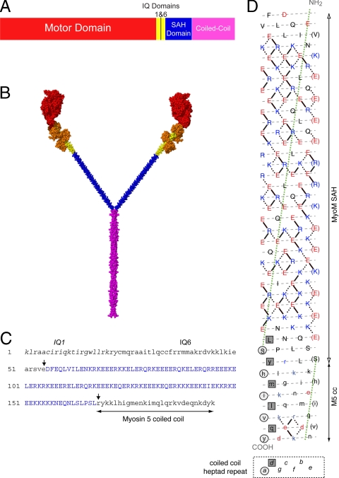
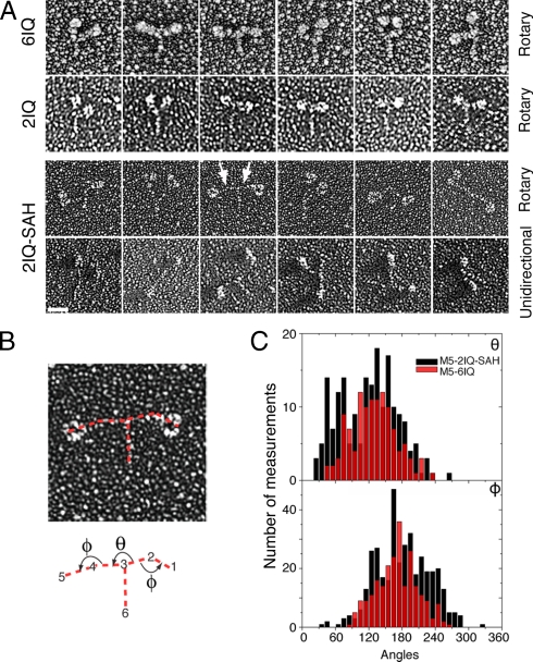
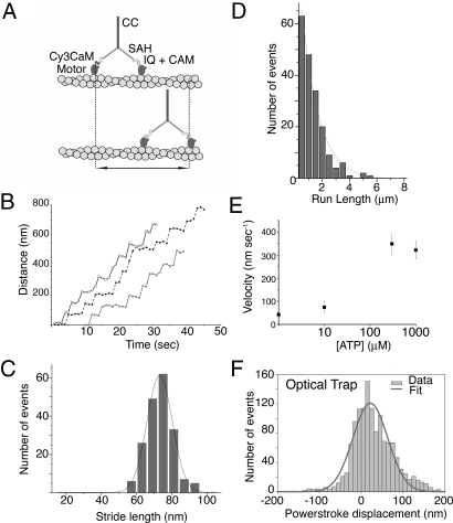
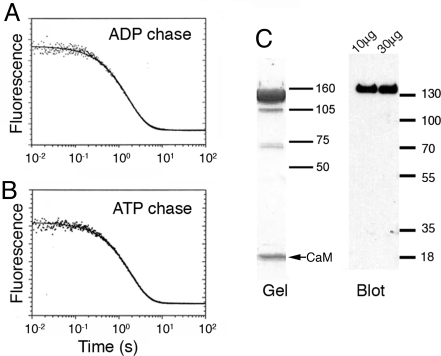
Stable, single alpha-helix (SAH) domains are widely distributed in the proteome, including in myosins, but their functions are unknown. To test whether SAH domains can act as levers, we replaced four of the six calmodulin-binding IQ motifs in the levers of mouse myosin 5a (Myo5) with the putative SAH domain of Dictyostelium myosin MyoM of similar length. The SAH domain was inserted between the IQ motifs and the coiled coil in a Myo5 HMM construct in which the levers were truncated from six to two IQ motifs (Myo5-2IQ). Electron microscopy of this chimera (Myo5-2IQ-SAH) showed the SAH domain was straight and 17 nm long as predicted, restoring the truncated lever to the length of wild-type (Myo5-6IQ). The powerstroke (of 21.5 nm) measured in the optical trap was slightly less than that for Myo5-6IQ but much greater than for Myo5-2IQ. Myo5-2IQ-SAH moved processively along actin at physiological ATP concentrations with similar stride and run lengths to Myo5-6IQ in in-vitro single molecule assays. In comparison, Myo5-2IQ is not processive under these conditions. Solution biochemical experiments indicated that the rear head did not mechanically gate the rate of ADP release from the lead head, unlike Myo5-6IQ. These data show that the SAH domain can form part of a functional lever in myosins, although its mechanical stiffness might be lower. More generally, we conclude that SAH domains can act as stiff structural extensions in aqueous solution and this structural role may be important in other proteins.


Conflict of interest statement

The authors declare no conflict of interest.

Figures

Fig. 1.
Fig. 1.
Diagram of the Myo5–2IQ-SAH chimera and the SAH domain of MyoM. (A) The domain arrangement of the chimeric Myo5–2IQ-SAH HMM construct. (B) An atomic model of the chimera molecule shown in spacefill using the same color scheme as in (A) with the addition of bound CaM molecules shown in orange. The construction of this model is described in SI Text. (C) The sequence in the chimera from the start of IQ1 to the start of the coiled-coil domain in Myo5a, to show the boundaries (arrowed) between Myo5a sequence (lowercase) and the inserted MyoM SAH domain (blue, uppercase). The original Myo5–2IQ construct used to generate the chimera has been described in ref. . (D) A heptad net representation of the MyoM SAH domain (uppercase) and the first 27 residues of the coiled coil of Myo5 HMM (lowercase). The helix has been cut beside one of the seven 18/1 helices (that would be designated a-g in an α-helical coiled coil) and opened flat to give a 2-D representation of the amino acid residues and their potential interactions in the helix, the N terminus at the top; acidic residues are shown in red, basic in blue, all others in black. The preferred ionic interactions (see ref. 2) are shown as solid lines. In addition alternative/other interactions are shown as dotted lines. Every seventh residue is repeated on the right of the plot (in brackets) so that all interactions can be shown. The gray dashed lines indicate the path of the polypeptide backbone; the green dotted line defines the orientation of the α-helix axis. The light gray circles indicate “a” positions and the dark gray squares indicate “d” positions that form the hydrophobic seam in the coiled coil of myosin 5. The last few residues of the SAH domain (uppercase) could, in principle, form a coiled coil in register with the coiled coil of Myo5 HMM, but there is no hydrophobic seam in the majority of the SAH domain, suggesting that it would not form coiled coil.
Fig. 2.
Fig. 2.
Electron Micrographs and analysis of rotary shadowed molecules of the Myo5–2IQ-SAH chimera. (A) A gallery of single molecules of rotary shadowed Myo5–6IQ and Myo5–2IQ, together with those for GFP-Myo5–2IQ-SAH HMM chimera as seen in either rotary or unidirectional shadowing. The arrows indicate the positions of the SAH domains in a molecule of the chimera. (Scale bar, 25 nm.) (B) Diagram of the chimera showing how measurement of the angles between domains was carried out. Red dotted lines indicate the lines between which the angles were measured between the Myo5–2IQ head and the SAH domain (Φ), and between the SAH domains (θ). To measure the angles for each molecule, SPIDER was used to select the tip of the head, head-SAH junction, SAH-SAH junction and the end of the coiled coil in the order shown. The angles were always measured in the anticlockwise direction as indicated by the arrows. (C) Distribution of the angles for the chimera, as indicated in part (B) (black) and for Myo5–6IQ (red).
Fig. 3.
Fig. 3.
Stride length data for Myo5–2IQ-SAH chimera from TIRF microscopy and FIONA, and powerstroke measurements from optical trap assays. (A) Diagram showing the movement that is measured for singly labeled heads, in this assay. (B) Example traces of stride length data for molecules that are labeled by Cy3 calmodulin bound to a single head show that the Myo5–2IQ-SAH chimera is able to move processively along actin. Because movement is stochastic, velocities can vary significantly even within a single run. (C) Histogram of stride lengths fitted with a Gaussian distribution for Cy3 labeled chimera with a label on one head. The mean value is 70.3 ± 7.5 nm (n = 188). Experiments were performed using 300 nM ATP. (D) Run lengths at 1 mM ATP (40 mM KCl) for the Myo5–2IQ-SAH chimera. n = 188, and the run length constant is 1,214 ± 52 nm. The line is an exponential fit to the data. (E) The ATP dependence of velocity of the Myo5–2IQ-SAH chimera on actin in TIRFM assays. n = 40–80 for each measurement. (F) The distribution of powerstroke displacements measured for Myo5–2IQ-SAH chimera in the optical trap, fitted by a Gaussian distribution with a mean value of 21.5 nm. The stiffness of the trap (0.015–0.030 pN·nm−1) implies the stroke is developed against a force of 0.3–0.7 pN. The breadth (S.E.M., 2.0 nm) largely derives from the imposed sine-wave oscillation imposed on the actin filament (see Methods).
Fig. 4.
Fig. 4.
Product dissociation from the acto-Myo5–2IQ-SAH-ADP-Pi complexes. (A) Myo5–2IQ-SAH chimera (0.4 μM) was mixed with 0.5 μM deac-aminoATP, held for 20 s in a delay line, then mixed with hexokinase-glucose treated phalloidin-actin and 2 mM ADP. Experimental conditions: 40 mM KCl, 10 mM Mops, 3 mM MgCl2, 1 mM EGTA, and 1 mM DTT, pH 7.5, 20 °C. Final concentrations in the cell: 0.11 μM Myo5–2IQ-SAH active sites, 0.14 μM deac-aminoATP, 11.1 μM actin, and 1.1 mM ADP. The solid line through the data are the best fit to a single exponential equation: I(t) = 0.079e−0.58t + C. (B) Experimental conditions were similar to those in (A) except that a 2 mM ATP chase replaced ADP: I(t) = 0.080e−0.52t + C. (C) SDS gel and Western blot (using an anti-FLAG antibody) of the Myo5–2IQ-SAH chimera used in these experiments. The major band on the gel and the blot shows that the purified protein mainly consists of full-length molecules. Two different loadings (10 μg and 30 μg) of protein were used in the blot. (Size markers in kDa.)
Fig. 5.
Fig. 5.
Diagram of the principal states of the ATP hydrolysis pathway of actin-Myo5–2IQ-SAH. T = ATP, D = ADP, P = phosphate. In the levers, the 2 IQ motifs with bound calmodulins are shown as ellipses, and the SAH domain by a blue rod, which is shown curved where it is distorted by intramolecular stresses. In wild-type Myo5–6IQ, ADP dissociation from the lead head (converting state 2 to 6, rate constant k2,6) is much slower than from the trail head and the principal pathway is the processive mechanism (1→2→3→4→5→1; gray shaded area) where one step is made for each ATP molecule hydrolyzed. ADP dissociation from the trail head (2→3) is rate limiting and the other steps on the processive path are at least 10-fold faster. Side paths that might lead to dissociation of wild-type Myo5–6IQ occur only rarely because ADP dissociates mainly from the trail head; that is, k2,3 ≫ k2,6 and k3,4 ≫ k3,7. Runs terminate mainly by release of the attached head (5→9→10) during the search phase of the cycle (5→1). For the Myo5–2IQ-SAH chimera, ADP dissociation from the trail and lead heads occurs at the same rate (k2,3 ≈ k2,6) and state 2 thus proceeds equally to states 3 and 6. Dissociation of ADP from the lead head would mostly lead to a “futile cycle” (6→1→2; blue shaded area) in which the lead head would consume a molecule of ATP without producing movement along actin. Processivity would be decreased slightly by the dissociation (7→8) that would occur upon state 7 binding two molecules of ATP, but usually binding of ATP to state 7 would allow a return to state 3 of the cycle. Short dotted arrows between 2 and 6 and between 3 and 7 indicate the slower rate of release of ADP from the lead head of Myo5–6IQ, compared to the chimera (solid arrows). Note that the canonical two IQ lever of the lead head is shown as post-powerstroke in slates 2, 3, 4, 6, and 7, unlike in wild type Myo5-6IQ.

Similar articles

  • Neck length and processivity of myosin V.
    Sakamoto T, Wang F, Schmitz S, Xu Y, Xu Q, Molloy JE, Veigel C, Sellers JR. Sakamoto T, et al. J Biol Chem. 2003 Aug 1;278(31):29201-7. doi: 10.1074/jbc.M303662200. Epub 2003 May 11. J Biol Chem. 2003. PMID: 12740393
  • Role of the lever arm in the processive stepping of myosin V.
    Purcell TJ, Morris C, Spudich JA, Sweeney HL. Purcell TJ, et al. Proc Natl Acad Sci U S A. 2002 Oct 29;99(22):14159-64. doi: 10.1073/pnas.182539599. Epub 2002 Oct 17. Proc Natl Acad Sci U S A. 2002. PMID: 12386339 Free PMC article.
  • Influence of lever structure on myosin 5a walking.
    Oke OA, Burgess SA, Forgacs E, Knight PJ, Sakamoto T, Sellers JR, White H, Trinick J. Oke OA, et al. Proc Natl Acad Sci U S A. 2010 Feb 9;107(6):2509-14. doi: 10.1073/pnas.0906907107. Epub 2010 Jan 25. Proc Natl Acad Sci U S A. 2010. PMID: 20133809 Free PMC article.
  • Myosin learns to walk.
    Mehta A. Mehta A. J Cell Sci. 2001 Jun;114(Pt 11):1981-98. doi: 10.1242/jcs.114.11.1981. J Cell Sci. 2001. PMID: 11493635 Review.
  • Regulation of myosin 5a and myosin 7a.
    Siththanandan VB, Sellers JR. Siththanandan VB, et al. Biochem Soc Trans. 2011 Oct;39(5):1136-41. doi: 10.1042/BST0391136. Biochem Soc Trans. 2011. PMID: 21936778 Review.

Cited by

References

    1. Berg JS, Powell BC, Cheney RE. A millennial myosin census. Mol Biol Cell. 2001;12:780–794. - PMC - PubMed
    1. Peckham M, Knight PJ. When a predicted coiled coil is really a single α-helix, in myosins and other proteins. Soft Matter. 2009;5:2493–2503.
    1. Mehta AD, et al. Myosin-V is a processive actin-based motor. Nature. 1999;400:590–593. - PubMed
    1. Veigel C, Wang F, Bartoo ML, Sellers JR, Molloy JE. The gated gait of the processive molecular motor, myosin V. Nat Cell Biol. 2002;4:59–65. - PubMed
    1. De La Cruz EM, Wells AL, Rosenfeld SS, Ostap EM, Sweeney HL. The kinetic mechanism of myosin V. Proc Natl Acad Sci USA. 1999;96:13726–13731. - PMC - PubMed

Publication types

MeSH terms