

A photoactivatable green-fluorescent protein from the phylum Ctenophora
- PMID: 20018790
- PMCID: PMC2842807
- DOI: 10.1098/rspb.2009.1774
A photoactivatable green-fluorescent protein from the phylum Ctenophora
Retraction in
-
A photoactivatable green-fluorescent protein from the phylum Ctenophora.Proc Biol Sci. 2015 Aug 7;282(1812):20151055. doi: 10.1098/rspb.2015.1055. Proc Biol Sci. 2015. PMID: 26224709 Free PMC article. No abstract available.
Abstract
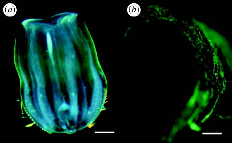
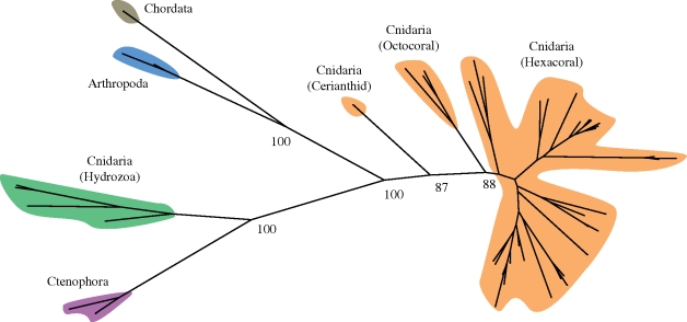
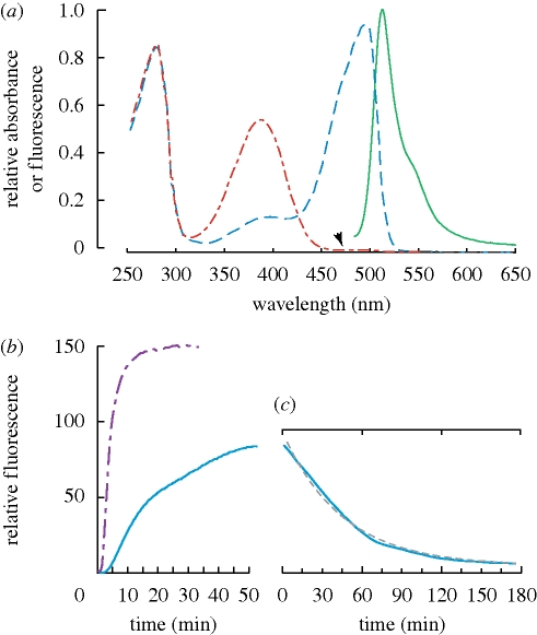
Genes for the family of green-fluorescent proteins (GFPs) have been found in more than 100 species of animals, with some species containing six or more copies producing a variety of colours. Thus far, however, these species have all been within three phyla: Cnidaria, Arthropoda and Chordata. We have discovered GFP-type fluorescent proteins in the phylum Ctenophora, the comb jellies. The ctenophore proteins share the xYG chromophore motif of all other characterized GFP-type proteins. These proteins exhibit the uncommon property of reversible photoactivation, in which fluorescent emission becomes brighter upon exposure to light, then gradually decays to a non-fluorescent state. In addition to providing potentially useful optical probes with novel properties, finding a fluorescent protein in one of the earliest diverging metazoans adds further support to the possibility that these genes are likely to occur throughout animals.
Figures
Similar articles
-
Genomic organization, evolution, and expression of photoprotein and opsin genes in Mnemiopsis leidyi: a new view of ctenophore photocytes.BMC Biol. 2012 Dec 21;10:107. doi: 10.1186/1741-7007-10-107. BMC Biol. 2012. PMID: 23259493 Free PMC article.
-
Extensive mitochondrial gene rearrangements in Ctenophora: insights from benthic Platyctenida.BMC Evol Biol. 2018 Apr 27;18(1):65. doi: 10.1186/s12862-018-1186-1. BMC Evol Biol. 2018. PMID: 29703131 Free PMC article.
-
Expression and characterization of the calcium-activated photoprotein from the ctenophore Bathocyroe fosteri: insights into light-sensitive photoproteins.Biochem Biophys Res Commun. 2013 Feb 8;431(2):360-6. doi: 10.1016/j.bbrc.2012.12.026. Epub 2012 Dec 19. Biochem Biophys Res Commun. 2013. PMID: 23262181 Free PMC article.
-
Green fluorescent protein.Photochem Photobiol. 1995 Oct;62(4):651-6. doi: 10.1111/j.1751-1097.1995.tb08712.x. Photochem Photobiol. 1995. PMID: 7480149 Review.
-
Evolutionary origin of the nervous system from Ctenophora prospective.Evol Dev. 2024 Jul;26(4):e12472. doi: 10.1111/ede.12472. Epub 2024 Feb 23. Evol Dev. 2024. PMID: 38390763 Review.
Cited by
-
Shining in the dark: First record of biofluorescence in the seahorse Hippocampus reidi.PLoS One. 2019 Aug 8;14(8):e0220561. doi: 10.1371/journal.pone.0220561. eCollection 2019. PLoS One. 2019. PMID: 31393893 Free PMC article.
-
A putative chordate luciferase from a cosmopolitan tunicate indicates convergent bioluminescence evolution across phyla.Sci Rep. 2020 Oct 20;10(1):17724. doi: 10.1038/s41598-020-73446-w. Sci Rep. 2020. PMID: 33082360 Free PMC article.
-
The covert world of fish biofluorescence: a phylogenetically widespread and phenotypically variable phenomenon.PLoS One. 2014 Jan 8;9(1):e83259. doi: 10.1371/journal.pone.0083259. eCollection 2014. PLoS One. 2014. PMID: 24421880 Free PMC article.
-
Fluorescence as a means of colour signal enhancement.Philos Trans R Soc Lond B Biol Sci. 2017 Jul 5;372(1724):20160335. doi: 10.1098/rstb.2016.0335. Philos Trans R Soc Lond B Biol Sci. 2017. PMID: 28533452 Free PMC article. Review.
-
A photoactivatable green-fluorescent protein from the phylum Ctenophora.Proc Biol Sci. 2015 Aug 7;282(1812):20151055. doi: 10.1098/rspb.2015.1055. Proc Biol Sci. 2015. PMID: 26224709 Free PMC article. No abstract available.
References
-
- Alieva N. O., et al. 2008Diversity and evolution of coral fluorescent proteins. PLoS ONE 3, e2680 (doi:10.1371/journal.pone.0002680) - DOI - PMC - PubMed
-
- Ando R., Hama H., Yamamoto-Hino M., Mizuno H.2002An optical marker based on the UV-induced green-to-red photoconversion of a fluorescent protein. Proc. Natl Acad. Sci. USA 99, 12 651–12 656 (doi:10.1073/pnas.202320599) - DOI - PMC - PubMed
-
- Ando R., Mizuno H., Miyawaki A.2004Regulated fast nucleocytoplasmic shuttling observed by reversible protein highlighting. Science 306, 1370–1373 (doi:10.1126/science.1102506) - DOI - PubMed
-
- Barnes W. M.1994PCR amplification of up to 35-kb DNA with high fidelity and high yield from lambda bacteriophage templates. Proc. Natl Acad. Sci. USA 91, 2216–2220 (doi:10.1073/pnas.91.6.2216) - DOI - PMC - PubMed
-
- Bogdanov A. M., Mishin A. S., Yampolsky I. V., Belousov V. V., Chudakov D. M., Subach F. V., Verkhusha V. V., Lukyanov S., Lukyanov K. A.2009Green fluorescent proteins are light-induced electron donors. Nat. Chem. Biol. 5, 459–461 (doi:10.1038/nchembio.174) - DOI - PMC - PubMed
Publication types
MeSH terms
Substances
LinkOut - more resources
Full Text Sources
