

Role of neurotransmitter receptors in mediating light-evoked responses in retinal interplexiform cells
- PMID: 20018830
- PMCID: PMC2822699
- DOI: 10.1152/jn.00876.2009
Role of neurotransmitter receptors in mediating light-evoked responses in retinal interplexiform cells
Abstract
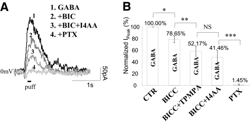
Interplexiform (IP) cells are a long-neglected group of retinal neurons the function of which is yet to be determined. Anatomical study indicates that IP cells are located in the inner nuclear layer, juxtaposed with the third-order neurons. However, the synaptic transmission of IP cells in the inner retina is poorly understood. Using whole cell patch-clamp and pharmacological techniques, we extensively studied synaptic receptors in IP cells. The IP cells in amphibian retinal slices were identified by electrical and morphological properties with voltage-clamp recording and Lucifer yellow dialysis. We find that light-evoked excitatory postsynaptic currents (L-EPSCs) are mediated by AMPA and N-methyl-d-aspartate receptors in IP cells. Although both receptors contributed to the amplitude and kinetics of L-EPSCs, AMPA receptor desensitization substantially shaped L-EPSCs in the neurons, similar to those found in the third-order neurons. The light-evoked inhibitory postsynaptic currents (L-IPSCs) in IP cells were primarily mediated by strychnine-sensitive glycine receptors with a small component of GABA(C) receptors. GABA(C) receptor rho2 subunits were detected in IP cells with single-cell RT-PCR assays. Expression of GABA(C) receptors is one of the special features for IP cells, distinct from most of the third-order neurons that depend on GABA(A) and glycine receptors to relay the inhibitory signals. However, GABA(A) receptors in IP cells acted like nonsynaptic receptors that were activated by exogenous GABA application. Furthermore, L-IPSCs in IP cells were inhibited by the serial inhibitions between amacrine cells in the inner retina. In addition, application of neurotransmitters on the axon terminals of IP cells had no significant current generated in the cells, indicating that the synaptic inputs of IP cells are mainly from the inner retina. This study demonstrates the important role that light signals are encoded by both experiment of inhibitory receptors in IP cells.
Figures


Similar articles
-
GABA(A), GABA(C) and glycine receptor-mediated inhibition differentially affects light-evoked signalling from mouse retinal rod bipolar cells.J Physiol. 2006 Apr 1;572(Pt 1):215-25. doi: 10.1113/jphysiol.2005.103648. Epub 2006 Jan 26. J Physiol. 2006. PMID: 16439422 Free PMC article.
-
Receptor and transmitter release properties set the time course of retinal inhibition.J Neurosci. 2006 Sep 13;26(37):9413-25. doi: 10.1523/JNEUROSCI.2591-06.2006. J Neurosci. 2006. PMID: 16971525 Free PMC article.
-
Multiple types of spontaneous excitatory synaptic currents in salamander retinal ganglion cells.Brain Res. 1999 Mar 13;821(2):487-502. doi: 10.1016/s0006-8993(99)01067-7. Brain Res. 1999. PMID: 10064836
-
AMPA-preferring receptors mediate excitatory synaptic inputs to retinal ganglion cells.J Neurophysiol. 1997 Jan;77(1):57-64. doi: 10.1152/jn.1997.77.1.57. J Neurophysiol. 1997. PMID: 9120596
-
Characterization of receptors for glutamate and GABA in retinal neurons.Prog Neurobiol. 2004 Jun;73(2):127-50. doi: 10.1016/j.pneurobio.2004.04.002. Prog Neurobiol. 2004. PMID: 15201037 Review.
Cited by
-
Retinal synaptic pathways underlying the response of the rabbit local edge detector.J Neurophysiol. 2010 May;103(5):2757-69. doi: 10.1152/jn.00987.2009. Epub 2010 Mar 24. J Neurophysiol. 2010. PMID: 20457864 Free PMC article.
-
Glycinergic feedback enhances synaptic gain in the distal retina.J Physiol. 2014 Apr 1;592(7):1479-92. doi: 10.1113/jphysiol.2013.265785. Epub 2014 Jan 13. J Physiol. 2014. PMID: 24421349 Free PMC article.
-
Retinal amino acid neurochemistry of the southern hemisphere lamprey, Geotria australis.PLoS One. 2013;8(3):e58406. doi: 10.1371/journal.pone.0058406. Epub 2013 Mar 13. PLoS One. 2013. PMID: 23516473 Free PMC article.
References
-
- Borges S, Wilson M. Dual effect of glycine on horizontal cells of the tiger salamander retina. J Neurophysiol 66: 1993–2001, 1991 - PubMed
-
- Boycott BB, Dowling JE, Fisher SK, Kolb H, Laties AM. Interplexiform cells of the mammalian retina and their comparison with catecholamine-containing retinal cells. Proc R Soc Lond B Biol Sci 191: 353–368, 1975 - PubMed
-
- Diamond JS, Copenhagen DR. The contribution of NMDA and non-NMDA receptors to the light-evoked input-output characteristics of retinal ganglion cells. Neuron 11: 725–738, 1993 - PubMed
-
- Djamgoz MBA, Laming PJ. Micro-electrode measurements and functional aspects of chloride activity in cyprinid fich retina: Extracellular activity and intracellular activities of L- and C-type horizontal cells. Vision Res 27: 1481–1489, 1987 - PubMed
Publication types
MeSH terms
Substances
Grants and funding
LinkOut - more resources
Full Text Sources