. 2010 Feb 12;285(7):4732-40.
doi: 10.1074/jbc.M109.086033. Epub 2009 Dec 17.

Identification of the minimal functional unit of the homo-oligomeric human reduced folate carrier

Affiliations

Identification of the minimal functional unit of the homo-oligomeric human reduced folate carrier

Zhanjun Hou et al. J Biol Chem. .

Abstract

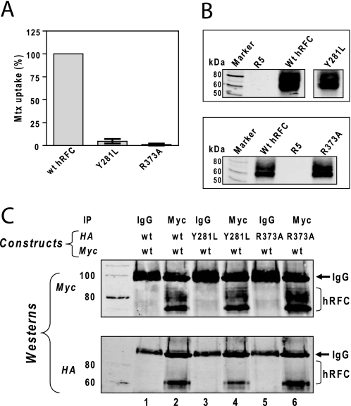
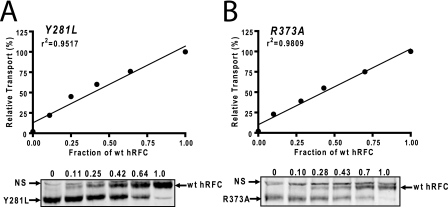
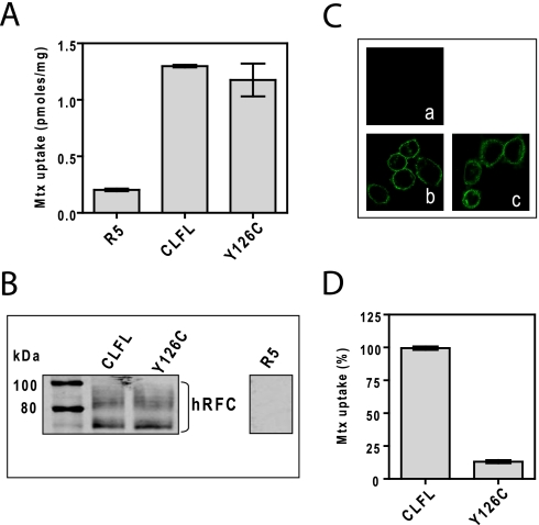
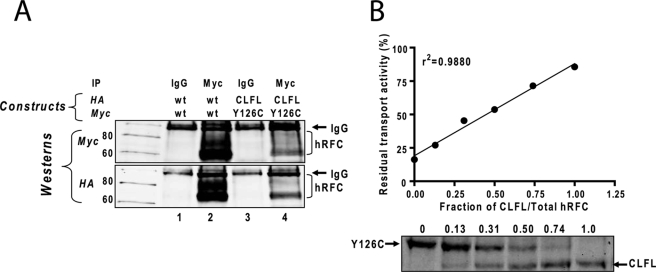
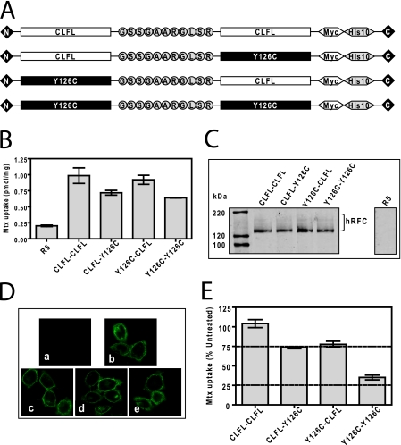
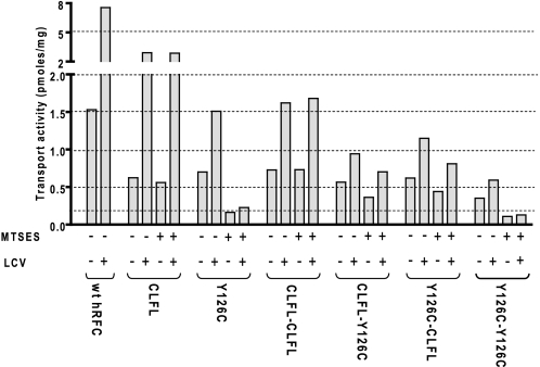
The reduced folate carrier (RFC) is the major transport system for folates in mammals. We previously demonstrated the existence of human RFC (hRFC) homo-oligomers and established the importance of these higher order structures to intracellular trafficking and carrier function. In this report, we examined the operational significance of hRFC oligomerization and the minimal functional unit for transport. In negative dominance experiments, multimeric transporters composed of different ratios of active (either wild type (WT) or cysteine-less (CLFL)) and inactive (either inherently inactive (Y281L and R373A) due to mutation, or resulting from inactivation of the Y126C mutant by (2-sulfonatoethyl) methanethiosulfonate (MTSES)) hRFC monomers were expressed in hRFC-null HeLa (R5) cells, and residual WT or CLFL activity was measured. In either case, residual transport activity with increasing levels of inactive mutant correlated linearly with the fraction of WT or CLFL hRFC in plasma membranes. When active covalent hRFC dimers, generated by fusing CLFL and Y126C monomers, were expressed in R5 cells and treated with MTSES, transport activity of the CLFL-CLFL dimer was unaffected, whereas Y126C-Y126C was potently (64%) inhibited; heterodimeric CLFL-Y126C and Y126C-CLFL were only partly (27 and 23%, respectively) inhibited by MTSES. In contrast to Y126C-Y126C, trans-stimulation of methotrexate uptake by intracellular folates for Y126C-CLFL and CLFL-Y126C was nominally affected by MTSES. Collectively, these results strongly support the notion that each hRFC monomer comprises a single translocation pathway for anionic folate substrates and functions independently of other monomers (i.e. despite an oligomeric structure, hRFC functions as a monomer).


Figures

FIGURE 1.
FIGURE 1.
Function and expression of inactive hRFC mutants and their association with WT hRFC. A and B, HA-tagged hRFC WT and its mutants (Y281L and R373A) were transiently transfected into HeLa R5 cells. A, cells were assayed for transport with [3H]Mtx (0.5 μm) over 2 min at 37 °C. Transport results are expressed as the averages ± ranges for two separate experiments. B, hRFC expression is shown on a Western blot of plasma membrane proteins (2.5 μg) from hRFC-null R5 cells, R5 transfectants expressing WT and Y281L hRFCs (top), and WT and R373A hRFCs (bottom). Detection of immunoreactive hRFC was with anti-HA primary antibody and IRDye800-conjugated secondary antibody and used an Odyssey® infrared imaging system. The molecular mass markers for SDS-PAGE (in kDa) are noted. C, Myc-tagged WT hRFC was co-expressed with HA-tagged WT, Y281L, or R373A hRFC in HeLa R5 cells. Plasma membranes were prepared, solubilized, and immunoprecipitated (IP) with Myc-specific antibody (lanes 2, 4, and 6) or with control IgG (lanes 1, 3, and 5). The samples were analyzed by SDS-PAGE and immunoblotted with Myc- and HA-specific antibodies. The molecular mass markers (in kDa) for SDS-PAGE are noted.
FIGURE 2.
FIGURE 2.
Determination of the minimal functional unit of hRFC by co-expressing WT and inactive hRFC mutants. WT (Myc-tagged) and mutant (Y281L and R373A; HA-tagged) hRFC constructs were transiently transfected into HeLa R5 cells. The cells were assayed for transport with [3H]Mtx (0.5 μm) over 2 min at 37 °C. Surface hRFC proteins labeled with sulfo-NHS-SS-biotin were measured by SDS-PAGE and Western blotting with anti-hRFC antibody. Relative levels of WT and mutant hRFC proteins were measured by densitometry with Odyssey® software. The calculated fractions of WT hRFC to total hRFC protein from each sample are noted above each lane. The residual transport activities from representative experiments were plotted versus WT hRFC fractions from densitometry measurements, in the presence of Y281L (A) or R373A hRFCs (B). Correlation coefficients were calculated by Prism software (version 4.0). Results are shown for a representative experiment. In replicate experiments, transport activity and densitometry values did not vary more than 10%. NS, nonspecific. Detailed methods are described under “Materials and Methods.”
FIGURE 3.
FIGURE 3.
Characterization of CLFL hRFC and a single Cys insertion mutant, Y126C hRFC. Both Myc-tagged CLFL hRFC and Y126C hRFC constructs were transiently transfected into HeLa R5 cells. A, cells were assayed for transport with [3H]Mtx (0.5 μm) over 2 min at 37 °C. Transport results are expressed as the averages ± ranges for two separate experiments. B, hRFC expression is shown on a Western blot of plasma membrane proteins (5 μg) from hRFC-null R5 cells, and R5 transfectants expressing CLFL hRFC or Y126C hRFC. Detection of immunoreactive hRFC was with anti-Myc antibody and IRDye800-conjugated secondary antibody with an Odyssey® infrared imaging system. The molecular mass markers for SDS-PAGE (in kDa) are noted. C, confocal results are shown for hRFC-null R5 cells (a) and for R5 transfectants expressing CLFL hRFC (b) or Y126C hRFC (c). The cells were fixed with 3.3% paraformaldehyde, permeabilized with 0.1% Triton X-100, and stained with anti-Myc primary antibody and Alexa Fluor® 488-conjugated secondary antibody. The slides were visualized with a Zeiss laser-scanning microscope model 510 using a ×63 water immersion lens. D, R5 cells expressing CLFL hRFC and Y126C hRFC were preincubated with and without 10 mm MTSES for 15 min at 37 °C. Cells were washed, and [3H]Mtx (0.5 μm) uptake was assayed at 37 °C for 2 min. Uptake is presented as a percentage of the level measured in the absence of MTSES. All transport results are expressed as average values ± ranges for two separate experiments.
FIGURE 4.
FIGURE 4.
Determination of the hRFC minimal functional unit by co-expressing CLFL and Y126C hRFC Cys insertion mutant. A, results of a co-immunoprecipitation (IP) of CLFL hRFC with Y126C hRFC are shown. HeLa R5 cells were cotransfected together with WT hRFC (Myc-tagged) and WT hRFC (HA-tagged) or with CLFL hRFC (HA-tagged) and Y126C hRFC (Myc-tagged). Plasma membranes were prepared, solubilized, and immunoprecipitated with Myc-specific antibody (lanes 2 and 4) or with control IgG (lanes 1 and 3). The samples were analyzed by SDS-PAGE and immunoblotted with Myc- and HA-specific antibodies. Molecular mass markers for SDS-PAGE (in kDa) are noted. B, both CLFL hRFC (HA-tagged) and Y126C hRFC (Myc-tagged) constructs were transiently transfected into HeLa R5 cells. The cells were assayed for transport with [3H]Mtx (0.5 μm) over 2 min at 37 °C. Surface hRFC proteins were biotinylated with sulfo-NHS-SS-biotin and were measured by SDS-PAGE/Western blotting with anti-hRFC antibody. Results are shown for levels of CLFL and Y126C hRFC proteins determined by densitometry, and the calculated fractions of CLFL hRFC to total hRFC proteins are noted above each lane. The residual transport activities after treatment with MTSES (10 mm) were plotted versus the fraction of CLFL hRFC measured by densitometry. Results are shown for a representative experiment. In replicate experiments, both transport activity and densitometry values did not vary more than 10%. The correlation coefficient was calculated by Prism software (version 4.0). Detailed methods are described under “Materials and Methods.”
FIGURE 5.
FIGURE 5.
Characterization of covalent dimeric hRFC concatamers and treatment with MTSES to identify functional interactions between hRFC monomers. HeLa R5 cells were transiently transfected with CLFL-CLFL, CLFL-Y126C, Y126C-CLFL, and Y126C-Y126C concatameric hRFCs. A, schematic representation of hRFC concatamers composed of covalently linked CLFL and Y126C monomers (tagged with a Myc-His10) is shown. B, cells were assayed for transport with [3H]Mtx (0.5 μm) over 2 min at 37 °C. Transport results are expressed as the averages ± ranges for two separate experiments. C, hRFC expression is shown on a Western blot of plasma membrane proteins (5 μg) from hRFC-null R5 cells and R5 transfectants expressing CLFL-CLFL, CLFL-Y126C, Y126C-CLFL, and Y126C-Y126C concatameric hRFCs. Detection of immunoreactive hRFCs was with anti-Myc antibody and IRDye800-conjugated secondary antibody with an Odyssey® infrared imaging system. The molecular mass markers for SDS-PAGE (in kDa) are noted. D, confocal results are shown for hRFC-null R5 cells (a), R5 transfectants expressing CLFL-CLFL (b), CLFL-Y126C (c), Y126C-CLFL (d), and Y126C-Y126C concatameric hRFCs (e). The cells were fixed with 3.3% paraformaldehyde, permeabilized with 0.1% Triton X-100, and stained with anti-Myc primary antibody and Alexa Fluor® 488-conjugated secondary antibody. The slides were visualized with a Zeiss laser-scanning microscope 510 using a ×63 water immersion lens. E, R5 cells expressing CLFL-CLFL, CLFL-Y126C, Y126C-CLFL, and Y126C-Y126C concatameric hRFCs were preincubated with and without 10 mm MTSES for 15 min at 37 °C. Cells were washed, and 0.5 μm [3H]Mtx uptake was assayed at 37 °C for 2 min. Uptake is presented as a percentage of the level measured in the absence of MTSES in excess of the residual transport in untransfected R5 cells (not shown). All transport results are expressed as average values ± ranges for two separate experiments. Details are provided under “Materials and Methods.”
FIGURE 6.
FIGURE 6.
Trans-stimulation of [3H]Mtx influx by leucovorin with monomeric and concatameric hRFCs. HeLa R5 cells were transiently transfected with WT, CLFL, Y126C, CLFL-CLFL, CLFL-Y126C, Y126C-CLFL, and Y126C-Y126C hRFCs. For MTSES-treated samples, transfected cells were pretreated with 10 mm MTSES for 15 min at 37 °C and then assayed for trans-stimulation. For trans-stimulation, the transfected cells were preincubated with or without 500 μm leucovorin (LCV) for 15 min at 37 °C, washed, and assayed for [3H]Mtx (0.5 μm) uptake. Details are provided under “Materials and Methods.” Uptake is presented as a percentage of the level measured in excess of the residual low transport in untransfected R5 cells. Data are from a single representative experiment performed twice with nearly identical results.

Similar articles

Cited by

References

    1. Stokstad E. L. R. (1990) in Folic Acid Metabolism in Health and Disease (Picciano M. F., Stokstad E. L. R., Greogory J. F. ed) pp. 1–21, Wiley-Liss, New York
    1. Sirotnak F. M., Tolner B. (1999) Annu. Rev. Nutr. 19, 91–122 - PubMed
    1. Matherly L. H., Goldman D. I. (2003) Vitam. Horm. 66, 403–456 - PubMed
    1. Matherly L. H., Hou Z., Deng Y. (2007) Cancer Metastasis Rev. 26, 111–128 - PubMed
    1. Goldman I. D. (1971) Ann. N.Y. Acad. Sci. 186, 400–422 - PubMed

Publication types

MeSH terms

Substances

LinkOut - more resources