

JNK pathway-associated phosphatase dephosphorylates focal adhesion kinase and suppresses cell migration
- PMID: 20018849
- PMCID: PMC2820775
- DOI: 10.1074/jbc.M109.060186
JNK pathway-associated phosphatase dephosphorylates focal adhesion kinase and suppresses cell migration
Abstract
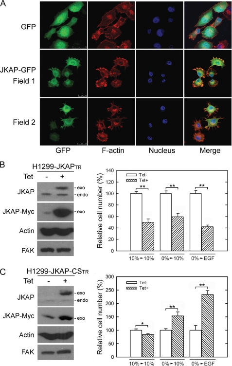
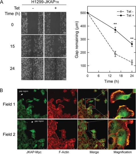
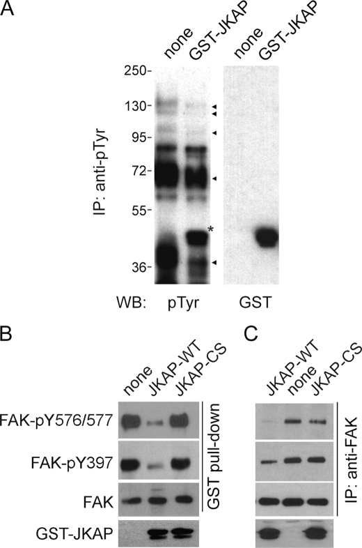
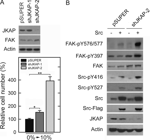
JNK pathway-associated phosphatase (JKAP, also named DUSP22) is expressed in various tissues, indicating that JKAP may have an important biological function. We showed that JKAP localized in the actin filament-enriched region. Expression of JKAP reduced cell migration, whereas a JKAP mutant lacking catalytic activity promoted cell motility. JKAP efficiently removed tyrosine phosphorylation of several proteins. We have identified focal adhesion kinase (FAK) as a substrate of JKAP. Overexpression of JKAP, but not JKAP mutant lacking catalytic activity, decreased FAK phosphorylation at tyrosines 397, 576, and 577 in H1299 cells. Consistent with these results, decreasing JKAP expression by RNA interference promoted cell migration and Src-induced FAK phosphorylation. Taken together, this study identified a new role for JKAP in the modulation of FAK phosphorylation and cell motility.
Figures

Similar articles
-
JNK pathway-associated phosphatase associates with rheumatoid arthritis risk, disease activity, and its longitudinal elevation relates to etanercept treatment response.J Clin Lab Anal. 2021 Apr;35(4):e23709. doi: 10.1002/jcla.23709. Epub 2021 Feb 6. J Clin Lab Anal. 2021. PMID: 33547838 Free PMC article.
-
Hepatocyte phosphatase DUSP22 mitigates NASH-HCC progression by targeting FAK.Nat Commun. 2022 Oct 8;13(1):5945. doi: 10.1038/s41467-022-33493-5. Nat Commun. 2022. PMID: 36209205 Free PMC article.
-
Downregulation of the phosphatase JKAP/DUSP22 in T cells as a potential new biomarker of systemic lupus erythematosus nephritis.Oncotarget. 2016 Sep 6;7(36):57593-57605. doi: 10.18632/oncotarget.11419. Oncotarget. 2016. PMID: 27557500 Free PMC article.
-
Scaffold Role of DUSP22 in ASK1-MKK7-JNK Signaling Pathway.PLoS One. 2016 Oct 6;11(10):e0164259. doi: 10.1371/journal.pone.0164259. eCollection 2016. PLoS One. 2016. PMID: 27711255 Free PMC article.
-
Hydrogen peroxide activates focal adhesion kinase and c-Src by a phosphatidylinositol 3 kinase-dependent mechanism and promotes cell migration in Caco-2 cell monolayers.Am J Physiol Gastrointest Liver Physiol. 2010 Jul;299(1):G186-95. doi: 10.1152/ajpgi.00368.2009. Epub 2010 Apr 8. Am J Physiol Gastrointest Liver Physiol. 2010. PMID: 20378826 Free PMC article.
Cited by
-
MAP4K Family Kinases and DUSP Family Phosphatases in T-Cell Signaling and Systemic Lupus Erythematosus.Cells. 2019 Nov 13;8(11):1433. doi: 10.3390/cells8111433. Cells. 2019. PMID: 31766293 Free PMC article. Review.
-
JNK Pathway-Associated Phosphatase Deficiency Facilitates Atherosclerotic Progression by Inducing T-Helper 1 and 17 Polarization and Inflammation in an ERK- and NF-κB Pathway-Dependent Manner.J Atheroscler Thromb. 2024 Oct 1;31(10):1460-1478. doi: 10.5551/jat.64754. Epub 2024 May 24. J Atheroscler Thromb. 2024. PMID: 38797677 Free PMC article.
-
Expression analyses of Dusp22 (Dual-specificity phosphatase 22) in mouse tissues.Med Mol Morphol. 2018 Jun;51(2):111-117. doi: 10.1007/s00795-017-0178-3. Epub 2017 Dec 27. Med Mol Morphol. 2018. PMID: 29282540
-
Shear force-based genetic screen reveals negative regulators of cell adhesion and protrusive activity.Proc Natl Acad Sci U S A. 2017 Sep 12;114(37):E7727-E7736. doi: 10.1073/pnas.1616600114. Epub 2017 Aug 28. Proc Natl Acad Sci U S A. 2017. PMID: 28847951 Free PMC article.
-
JNK pathway-associated phosphatase associates with rheumatoid arthritis risk, disease activity, and its longitudinal elevation relates to etanercept treatment response.J Clin Lab Anal. 2021 Apr;35(4):e23709. doi: 10.1002/jcla.23709. Epub 2021 Feb 6. J Clin Lab Anal. 2021. PMID: 33547838 Free PMC article.
References
-
- Patterson K. I., Brummer T., O'Brien P. M., Daly R. J. (2009) Biochem. J. 418, 475–489 - PubMed
-
- Barford D., Flint A. J., Tonks N. K. (1994) Science 263, 1397–1404 - PubMed
-
- Neel B. G., Tonks N. K. (1997) Curr. Opin. Cell Biol. 9, 193–204 - PubMed
-
- Alonso A., Sasin J., Bottini N., Friedberg I., Friedberg I., Osterman A., Godzik A., Hunter T., Dixon J., Mustelin T. (2004) Cell 117, 699–711 - PubMed
-
- Chen A. J., Zhou G., Juan T., Colicos S. M., Cannon J. P., Cabriera-Hansen M., Meyer C. F., Jurecic R., Copeland N. G., Gilbert D. J., Jenkins N. A., Fletcher F., Tan T. H., Belmont J. W. (2002) J. Biol. Chem. 277, 36592–36601 - PubMed
Publication types
MeSH terms
Substances
Grants and funding
LinkOut - more resources
Full Text Sources
Other Literature Sources
Molecular Biology Databases
Research Materials
Miscellaneous
