. 2010 Feb 19;285(8):5630-8.
doi: 10.1074/jbc.M109.074393. Epub 2009 Dec 14.

Involvement of the spliceosomal U4 small nuclear RNA in heterochromatic gene silencing at fission yeast centromeres

Affiliations

Involvement of the spliceosomal U4 small nuclear RNA in heterochromatic gene silencing at fission yeast centromeres

Madoka Chinen et al. J Biol Chem. .

Abstract

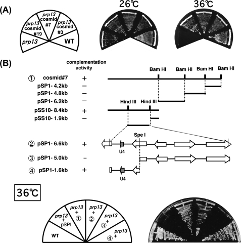
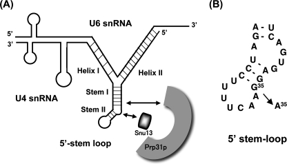
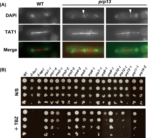
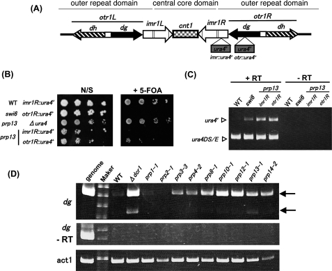
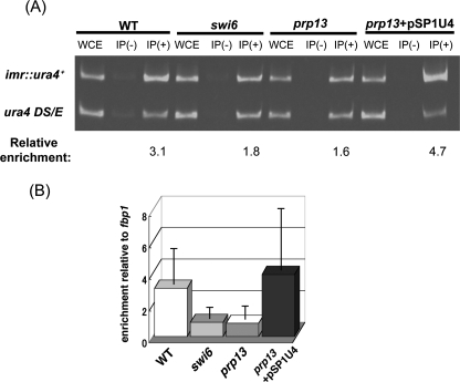
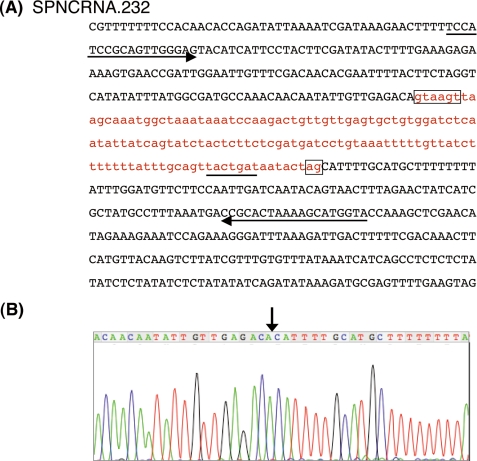
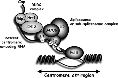
prp13-1 is one of the mutants isolated in a screen for defective pre-mRNA splicing at a nonpermissive temperature in fission yeast Schizosaccharomyces pombe. We cloned the prp13(+) gene and found that it encodes U4 small nuclear RNA (snRNA) involved in the assembly of the spliceosome. The prp13-1 mutant produced elongated cells, a phenotype similar to cell division cycle mutants, and displays a high incidence of lagging chromosomes on anaphase spindles. The mutant is hypersensitive to the microtubule-destabilizing drug thiabendazole, supporting that prp13-1 has a defect in chromosomal segregation. We found that the prp13-1 mutation resulted in expression of the ura4(+) gene inserted in the pericentromeric heterochromatin region and reduced recruitment of the heterochromatin protein Swi6p to that region, indicating defects in the formation of pericentromeric heterochromatin, which is essential for the segregation of chromosomes, in prp13-1. The formation of centromeric heterochromatin is induced by the RNA interference (RNAi) system in S. pombe. In prp13-1, the processing of centromeric noncoding RNAs to siRNAs, which direct the heterochromatin formation, was impaired and unprocessed noncoding RNAs were accumulated. These results suggest that U4 snRNA is required for the RNAi-directed heterochromatic gene silencing at the centromeres. In relation to the linkage between the spliceosomal U4 snRNA and the RNAi-directed formation of heterochromatin, we identified a mRNA-type intron in the centromeric noncoding RNAs. We propose a model in which the assembly of the spliceosome or a sub-spliceosome complex on the intron-containing centromeric noncoding RNAs facilitates the RNAi-directed formation of heterochromatin at centromeres, through interaction with the RNA-directed RNA polymerase complex.


Figures

FIGURE 1.
FIGURE 1.
Cloning of the prp13+ gene. A, complementation of the temperature-sensitive phenotype of prp13-1. Cosmids 3, 7, and 19 complemented the temperature-sensitive phenotype of prp13-1 at 36 °C. B, three DNA fragments were isolated from cosmid 7 after digestion with BamHI and subcloned into the pSPI vector (pSPI-4.2 kb, -4.8 kb, and -6.2 kb). The 8.4-kb fragment is a self-ligated clone of the BamHI-digested cosmid 7 (pSS10-8.4 kb). All subclones were introduced into prp13-1 and tested for complementation. Only the 8.4-kb self-ligated fragment complemented the prp13-1 mutation. Subcloning of the restriction fragments from pSS10-8.4 kb revealed that the 1.6-kb fragment could rescue the prp13-1 mutation. The 1.6-kb fragment contains the entire coding region for U4 snRNA.
FIGURE 2.
FIGURE 2.
Schematic representation of the U4/U6 structure and the mutation site in prp13-1. A, structure of the U4/U6 snRNA. U4 snRNA base pairs with U6 snRNA and has a 5′ stem-loop (5′ SL) structure (27). Snu13p and Prp31p bind directly with the 5′ SL region of U4 snRNA (38). B, in prp13-1, G35 in the 5′ SL region of U4 snRNA is changed to A.
FIGURE 3.
FIGURE 3.
prp13-1 yields lagging chromosomes and is sensitive to TBZ treatment. A, mitotic chromosomal segregation is impaired in prp13-1, resulting in a high incidence of lagging chromosomes during late anaphase. The prp13-1 cells were double stained with DAPI that binds DNA and the TAT1 antibody that binds tubulin. In the merged images (Merge), green and red denote DNA and tubulin, respectively. Two independent prp13-1 cells cultured at the permissive temperature are shown. Arrowheads indicate the lagging chromosomes. B, sensitivity to TBZ of the prp mutants. Serially diluted cells were spotted on YEALU plates without TBZ (N/S), or with 10 μg/ml of TBZ (+TBZ). The plates were incubated at 26 °C for ts, or 33 °C for cs mutants (prp11-1, prp14-2). Δdcr1 and Δago1 were spotted as controls for defective chromosomal segregation.
FIGURE 4.
FIGURE 4.
The mutation in U4 snRNA results in defective gene silencing at the centromere. A, schematic representation of the structure of the fission yeast centromere 1. Vertical lines in the imr1L and imr1R regions indicate clusters of (or single) tRNA genes that have been proposed to function as boundary elements (39). The central core domain is comprised of cnt and the inner part of the imr elements. The outer repeat domain encompasses the dg/dh elements and a small part of the imr elements. The ura4+ marker gene was inserted in the imr1R or otr1R region. B, serially diluted cells containing the inserted ura4+ gene were spotted on YEALU plates (N/S), or YEALU plates containing 1 mg/ml of 5-FOA and incubated at 26 °C. prp13-1 with the ura4+ gene inserted in the imr1R or otr1R region was sensitive to 5-FOA, indicating that the ura4+ marker gene was expressed in these strains. The swi6 mutant containing the ura4+ transgene, the wild type of which encodes a chromodomain protein, was used as a control for defects in the formation of centromeric heterochromatin. C, expression of the ura4+ mRNAs from the genes inserted in the pericentromere region was confirmed by RT-PCR. Total RNA was isolated from indicated strains and subjected to a RT-PCR assay. Reverse transcription was done with an oligo dT primer. Ura4DS/E indicates bands amplified from the authentic gene with the 269-bp deletion. D, accumulation of the centromeric dg noncoding RNA in the prp mutants. The prp mutants were cultured at the permissive temperature of 26 °C. Total RNA was isolated from the indicated strains and subjected to RT-PCR using primers corresponding to the dg noncoding RNA or act1 mRNA. No bands were detected in any samples without the reverse transcription reaction (−RT). Act1 mRNA was amplified by RT-PCR as an internal control. Upper and lower arrows denote bands derived from the authentic dg noncoding RNA and its spliced form, respectively.
FIGURE 5.
FIGURE 5.
The prp13-1 mutation abolished heterochromatin modifications at the otr1R::ura4+ locus. A, ChIP analyses of Swi6p at otr1R::ura4+ relative to a euchromatic control locus (ura4 DS/E) were performed with the indicated strains. Relative enrichment was calculated as the ratio of otr1R::ura4+ to ura4 DS/E in the immunoprecipitate, IP(+), relative to whole cell extract, WCE. The average enrichment values for three independent immunoprecipitations are shown. The reduction in the recruitment of Swi6p to the otr1R::ura4+ locus in prp13-1 was recovered by transformation of the wild-type U4 snRNA gene (lanes in prp13+pSP1U4). A representative gel is shown. B, real-time quantitative PCR analyses were carried out with the samples analyzed in A to assess the Swi6p enrichment at the otr1R::ura4+ locus relative to that of the fbp1 euchromatic control locus. Error bars represent S.D.
FIGURE 6.
FIGURE 6.
Impaired pre-mRNA splicing is not a cause of defective centromeric silencing. A, prp13-1 shows weak or no defects in pre-mRNA splicing. Total RNA was isolated from strains cultured at 37 °C for the periods indicated and analyzed by RT-PCR using primers for tbp1 or cdc2. White and black arrowheads indicate bands for pre-mRNAs and mature mRNAs, respectively. The cdc2 gene has two introns. B, no severe defects were observed in the splicing of hrr1+, ago1+, and sir2+ pre-mRNAs, the products of which are essential for the RNAi pathway. Total RNA extracted from the wild-type 972 cells or prp13-1 cells cultured at 26 °C or 37 °C for 2 h was subjected to a RT-PCR assay using primers for the indicated genes. Ago1 pre-mRNA is spliced with a low efficiency in both the wild-type and prp13-1 cells. C, introduction of the genes for U4 snRNA, Prp31p, and Snu13p recovered the centromeric gene silencing in prp13-1. Each strain or transformant was spotted on YEALU plates with 5-FOA (+FOA) or without 5-FOA (N/S), and incubated at 26 °C for 5 days. Two independent clones (1 and 2) were spotted for each transformant.
FIGURE 7.
FIGURE 7.
The centromeric dg noncoding RNA contains an mRNA-type intron. A, nucleotide sequence of the region containing the mRNA-type intron in the centromeric dg element. A part of the genomic region, where the dg noncoding RNA is transcribed from, is shown. The intron region is written in red lowercase letters. The sequences that matched the consensus sequence of the 5′ and 3′ splice sites in S. pombe are boxed. The putative branch site is underlined. Arrows indicate primers used for the RT-PCR analysis. B, intron-like region in the dg noncoding RNA is precisely removed in the cDNA corresponding to the lower band observed in Fig. 4D. Sequence data determined using the ABI 310 sequencer are shown. A vertical arrow indicates the position of the intron-like sequence in the dg noncoding RNA.
FIGURE 8.
FIGURE 8.
A hypothetical model for the involvement of the spliceosomal components in the RNAi-directed formation of heterochromatin. A specific set of splicing factors, including the U4/U6 snRNP, or ordinal spliceosomal components are assembled on the mRNA-type intron in the nascent noncoding RNAs transcribed from the pericentromeric otr region. Using the assembled sub-spliceosome complex or spliceosome as a platform, RDRC is recruited to the noncoding RNAs through interaction between Cid12p in RDRC and splicing factors, such as Cwf10p. RDRC then converts the centromeric noncoding RNAs into the double-stranded RNAs that are processed into siRNAs by Dicer.

Similar articles

Cited by

References

    1. Jurica M. S., Moore M. J. (2003) Mol. Cell 12, 5–14 - PubMed
    1. Burge C. B., Tuschl T., Sharp P. A. (1999) in The RNA Word (Gesteland R. F., Cech T. R., Atkins J. F. eds) pp. 525–560, Cold Spring Harbor Laboratory Press, Cold Spring Harbor, New York
    1. Valadkhan S. (2005) Curr. Opin. Chem. Biol. 9, 603–608 - PubMed
    1. Staley J. P., Guthrie C. (1998) Cell 92, 315–326 - PubMed
    1. Potashkin J., Li R., Frendewey D. (1989) EMBO J. 8, 551–559 - PMC - PubMed

Publication types

MeSH terms