. 2010 Feb 19;285(8):5479-87.
doi: 10.1074/jbc.M109.075606. Epub 2009 Dec 14.

NF-kappaB functions in stromal fibroblasts to regulate early postnatal muscle development

Affiliations

NF-kappaB functions in stromal fibroblasts to regulate early postnatal muscle development

Jason M Dahlman et al. J Biol Chem. .

Abstract

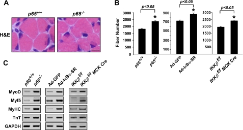
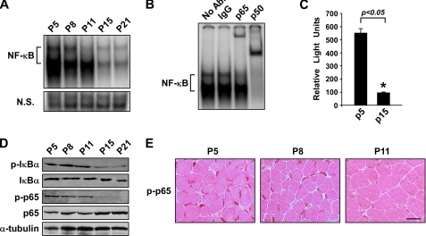
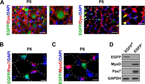
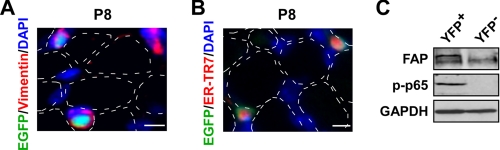
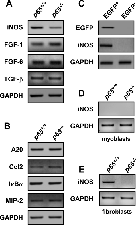
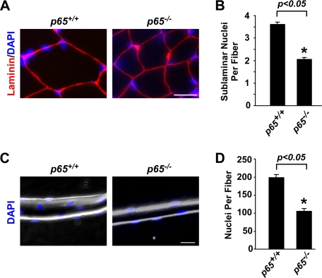
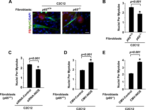
Classical NF-kappaB activity functions as an inhibitor of the skeletal muscle myogenic program. Recent findings reveal that even in newborn RelA/p65(-/-) mice, myofiber numbers are increased over that of wild type mice, suggesting that NF-kappaB may be a contributing factor in early postnatal skeletal muscle development. Here we show that in addition to p65 deficiency, repression of NF-kappaB with the IkappaB alpha-SR transdominant inhibitor or with muscle-specific deletion of IKKbeta resulted in similar increases in total fiber numbers as well as an up-regulation of myogenic gene products. Upon further characterization of early postnatal muscle, we observed that NF-kappaB activity progressively declines within the first few weeks of development. At birth, the majority of this activity is compartmentalized to muscle fibers, but by neonatal day 8 NF-kappaB activity from the myofibers diminishes, and instead, stromal fibroblasts become the main cellular compartment within the muscle that contains active NF-kappaB. We find that NF-kappaB functions in these fibroblasts to regulate inducible nitric-oxide synthase expression, which we show is important for myoblast fusion during the growth and maturation process of skeletal muscle. Together, these data broaden our understanding of NF-kappaB during development by showing that in addition to its role as a negative regulator of myogenesis, NF-kappaB also regulates nitric-oxide synthase expression within stromal fibroblasts to stimulate myoblast fusion and muscle hypertrophy.


Figures

FIGURE 1.
FIGURE 1.
Classical NF-κB signaling functions as a myogenic inhibitor during postnatal muscle development. A, cross-sections of P8 hind limbs from p65+/+;TNF−/− (p65+/+) and p65−/−;TNF−/− (p65−/−) mice were stained with hematoxylin and eosin (H&E). Scale bar = 10 μm. B, soleus muscle fiber counts were performed on hind limb cross-sections of P8 p65+/+ and p65−/−, Ad-GFP and Ad-IκBα-SR, and IKKβf/f and IKKβf/f MCK-CRE mice (n = 3) as described under “Experimental Procedures.” C, mRNA was isolated from the skeletal muscles of p65+/+ and p65−/−, Ad-GFP and Ad-IκBα-SR, and IKKβf/f and IKKβf/f MCK-CRE mice, and expression of myogenic markers, MyoD, Myf5, MyHC, and troponin (TNT) were visualized by semiquantitative PCR. GAPDH, glyceraldehyde-3-phosphate dehydrogenase.
FIGURE 2.
FIGURE 2.
Classical NF-κB signaling activity is reduced during early postnatal skeletal muscle development. A, electrophoretic mobility shift assay analysis was performed on nuclear extracts isolated from the skeletal muscles of P5-P21 wild type mice. Equal loading of nuclear extracts was compared with a nonspecific band on the same gel (N.S.). B, electrophoretic mobility shift assay super shift analysis was performed on P5 nuclear extracts from A with antibodies raised against the p65 and p50 subunits. Ab, antibody. C, whole cell extracts were isolated from the skeletal muscles of P5 and P15 NF-κB reporter mice (3xκB-Luc-Tg mice) mice and subjected to luciferase assay analysis. D and E, whole cell lysates were prepared from wild type P5-P21 skeletal muscle in homogenizing buffer followed by Western blot analysis for p-IκBα and p-p65 (D) as well as immunohistochemical staining for p-p65 on wild type P5-P11 hind limb cross-sections (E). Eosin was used as a counterstain as shown in E. Scale bar = 20 μm.
FIGURE 3.
FIGURE 3.
NF-κB activity switches cellular compartments during postnatal skeletal muscle development. A, immunofluorescent staining for dystrophin (Dys; red) was performed on hind limb cross-sections of P0 and P8 NF-κB+/EGFP (EGFP; green) mice. Areas shown as dashed boxes represent fields magnified at 2×. Scale bars = 20 and 10 μm, respectively. B and C, P8 NF-κB+/EGFP (EGFP; green) hind limb muscle cross-sections were immunostained with Pax7 (red, B) or MyoD (red, C). Dotted outlines denote the relative position of myofibers that were identified by the overexposure of fluorescent and phase contrast imaging. Scale bar = 5 μm. D, mononuclear cells were prepared from P8 NF-κB+/EGFP mice. EGFP+ and EGFP cells were isolated by FACS sorting and further probed by semiquantitative PCR for EGFP, MyoD, and Pax7. GAPDH, glyceraldehyde-3-phosphate dehydrogenase.
FIGURE 4.
FIGURE 4.
NF-κB is activated in the stromal fibroblasts of postnatal skeletal muscle. A and B, immunofluorescent staining was performed on P8 NF-κB+/EGFP (EGFP; green) hind limb muscle cross-sections with the mesenchymal marker vimentin (red; A) and the fibroblast-specific marker ER-TR7 (red; B). Similar to Fig. 3, the dotted outlines indicate the relative position of myofibers. Scale bar = 5 μm. C, YFP+ and YFP mononuclear cells were isolated from P8 collagen type 1a YFP transgenic mice and FACS sorted. Whole cell lysates were prepared from these individual cellular populations and Western blots were performed probing for p-p65 and fibroblast-activating protein (FAP). GAPDH, glyceraldehyde-3-phosphate dehydrogenase.
FIGURE 5.
FIGURE 5.
NF-κB regulates iNOS expression in stromal fibroblasts during postnatal muscle development. A and B, mRNA was isolated from the skeletal muscles of P8 p65+/+ and p65−/− mice and the expression of iNOS, fibroblast growth factor-1 and -6, and TGF-β (A) as well as other (B) established NF-κB-regulated genes was examined by semiquantitative PCR. C, EGFP+ and EGFP mononuclear cells were sorted from NF-κB+/EGFP mice and probed for iNOS expression by semiquantitative PCR. D and E, primary myoblasts and fibroblasts were isolated from p65+/+and p65−/− mice and iNOS expression was determined by semiquantitative PCR. GAPDH, glyceraldehyde-3-phosphate dehydrogenase.
FIGURE 6.
FIGURE 6.
Loss of p65 leads to a decrease in myofiber nucleation. A and B, TA muscles from 4-week-old p65+/+ and p65−/− mice (n = 3) were sectioned and stained with laminin and DAPI. Histological analysis was subsequently performed to quantitate sublaminar nuclei per fiber numbers. Scale bar = 10 μm. C and D, single fibers were isolated from the gastrocnemius muscles of 4-week-old p65+/+ and p65−/− mice and then stained with DAPI to analyze the total number of nuclei per fiber by fluorescent and phase contrast microscopy. Scale bar = 20 μm. Quantitation for B and D was performed as described under “Experimental Procedures.”
FIGURE 7.
FIGURE 7.
iNOS expression regulates myoblast fusion. A and B, p65+/+ and p65−/− fibroblasts (FB; red) were stained with 20 μm Cell Tracker Orange CMTMR and co-cultured for 24 h with C2C12 myoblasts. After 24 h of co-culturing, cells were switched to differentiation media, and myogenesis proceeded for 3 days after which cells were fixed and stained with MyHC (green) and DAPI (blue). Nuclei per myotube numbers were determined as described under “Experimental Procedures.” Scale bar = 40 μm. C and D, a similar analysis as described above was performed using wild type MEFs transfected with control or iNOS siRNA oligomers (C) or with control or iNOS cytomegalovirus expression plasmids (D). E, a similar analysis as described in A was performed using p65−/− MEFs transfected with control or iNOS cytomegalovirus expression plasmids.
FIGURE 8.
FIGURE 8.
Loss of iNOS expression inhibits myoblast fusion. A, primary iNOS+/+ and iNOS−/− fibroblasts were stained with 20 μm Cell Tracker Orange CMTMR and co-cultured for 24 h with C2C12 myoblasts. Differentiation was allowed to proceed for 3 days after which cells were fixed and stained with MyHC and DAPI and subsequently scored for nuclei per myotube. B, TA muscles from 4-week-old iNOS+/+ and iNOS−/− mice (n = 3) were sectioned and stained with laminin and DAPI. Sublaminar nuclei per fiber counts were determined by fluorescent microscopy. C, single fibers isolated from the gastrocnemius muscles of 4-week-old iNOS+/+ and iNOS−/− mice were stained with DAPI and analyzed by fluorescent and phase contrast microscopy to quantitate nuclei per fiber. Quantitation for A–C was performed as described under “Experimental Procedures.”

Similar articles

Cited by

References

    1. Baeuerle P. A., Baltimore D. (1996) Cell 87, 13–20 - PubMed
    1. Baldwin A. S., Jr. (1996) Annu. Rev. Immunol. 14, 649–683 - PubMed
    1. Verma I. M., Stevenson J. K., Schwarz E. M., Van Antwerp D., Miyamoto S. (1995) Genes Dev. 9, 2723–2735 - PubMed
    1. Hayden M. S., Ghosh S. (2004) Genes Dev. 18, 2195–2224 - PubMed
    1. Li Z., Nabel G. J. (1997) Mol. Cell. Biol. 17, 6184–6190 - PMC - PubMed

Publication types

MeSH terms