. 2010 Feb 19;285(8):5738-47.
doi: 10.1074/jbc.M109.066910. Epub 2009 Dec 15.

An experimentally based computer search identifies unstructured membrane-binding sites in proteins: application to class I myosins, PAKS, and CARMIL

Affiliations

An experimentally based computer search identifies unstructured membrane-binding sites in proteins: application to class I myosins, PAKS, and CARMIL

Hanna Brzeska et al. J Biol Chem. .

Abstract

Programs exist for searching protein sequences for potential membrane-penetrating segments (hydrophobic regions) and for lipid-binding sites with highly defined tertiary structures, such as PH, FERM, C2, ENTH, and other domains. However, a rapidly growing number of membrane-associated proteins (including cytoskeletal proteins, kinases, GTP-binding proteins, and their effectors) bind lipids through less structured regions. Here, we describe the development and testing of a simple computer search program that identifies unstructured potential membrane-binding sites. Initially, we found that both basic and hydrophobic amino acids, irrespective of sequence, contribute to the binding to acidic phospholipid vesicles of synthetic peptides that correspond to the putative membrane-binding domains of Acanthamoeba class I myosins. Based on these results, we modified a hydrophobicity scale giving Arg- and Lys-positive, rather than negative, values. Using this basic and hydrophobic scale with a standard search algorithm, we successfully identified previously determined unstructured membrane-binding sites in all 16 proteins tested. Importantly, basic and hydrophobic searches identified previously unknown potential membrane-binding sites in class I myosins, PAKs and CARMIL (capping protein, Arp2/3, myosin I linker; a membrane-associated cytoskeletal scaffold protein), and synthetic peptides and protein domains containing these newly identified sites bound to acidic phospholipids in vitro.


Figures

FIGURE 1.
FIGURE 1.
Binding of dansyl-labeled synthetic AMIC(tail) peptide to acidic phospholipid vesicles. A, fluorescence spectra of dansyl-labeled synthetic peptide with the sequence of the BH region in the tail of AMIC (see Table 1) in the presence of buffer alone, or 500 μm phospholipid vesicles contain 100% PC, 100% PS or 50% PS and 50% PC. B, increase in fluorescence intensity of dansyl-AMIC(tail) peptide as a function of concentration of 50% PS vesicles. The open and closed symbols are two independent experiments. Excitation, 335 nm; emission, 515 nm.
FIGURE 2.
FIGURE 2.
Comparison of hydrophobicity plots and BH plot of the Acanthamoeba myosin IC heavy chain. The amino acid values for the five different scales are given in Table 2. A window size of 19 was used for all plots.
FIGURE 3.
FIGURE 3.
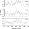
Comparison of BH plots of heavy chains of Acanthamoeba myosins IA, IB, and IC.
FIGURE 4.
FIGURE 4.
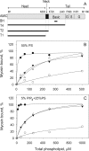
Binding of truncated Acanthamoeba myosin IC constructs to acidic phospholipid vesicles. A, schematic representation of the structure of the AMC heavy chain and the truncated constructs T4, T2, and T1. The head domain contains the actin-activated ATPase site. The neck domain contains the binding site for a single light chain. The tail domain contains an N-terminal basic region, two Gly/Pro/Ala-rich regions (G), and an SH3 domain (S). The BH region (bar) is located within the basic region. T4 comprises the head, neck and basic regions; T2 comprises the head and neck regions, and T1 is the head region only. B and C, binding of AMIC constructs to 50% PS vesicles (B) and 5% PIP2 and 50% PS vesicles (C). Binding was assayed by pelleting the vesicles assay and determining the percentage of unbound ATPase activity in the supernatant (see “Experimental Procedures”). T4 (filled circles) was coexpressed and purified with light chain, T2 both with (filled triangles) with and without (open triangles) light chain, and T1 (open circles) without light chain.
FIGURE 5.
FIGURE 5.
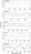
BH plots of proteins with known membrane-binding sites. N-WASP, EGFR, PLCζ1, and myosin VI were run with window size 19, and gelsolin was run with with window size 11.
FIGURE 6.
FIGURE 6.
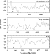
BH plots of Acanthamoeba, Dictyostelium, and mammalian PAKs. The positions of the CRIB domains (*) are Acanthamoeba PAK-(93–106), Dictyostelium PAK-(356–369), and mammalian PAK-(75–88).
FIGURE 7.
FIGURE 7.
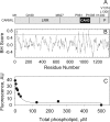
BH plot and phospholipid-binding of CARMIL. A, schematic representation of domain structure of mouse CARMIL 1: leucine-rich repeat (LRR); CARMIL homology domain 3, which binds actin capping protein, (CAH3); proline-rich region (P). B, BH plot with window size 19. C, binding of GFP-CAH3 to 50% PS vesicles. CAH3 was expressed with N-terminal GFP. Binding was assayed by pelleting the vesicles and measuring GFP fluorescence of the supernatant (see “Experimental Procedures”).

References

    1. Lemmon M. A. (2008) Nat. Rev. Mol. Cell Biol. 9, 99–111 - PubMed
    1. Niggli V. (2001) Trends Biochem. Sci. 26, 604–611 - PubMed
    1. Brzeska H., Hwang K. J., Korn E. D. (2008) J. Biol. Chem. 283, 32014–32023 - PMC - PubMed
    1. Lynch T. J., Brzeska H., Miyata H., Korn E. D. (1989) J. Biol. Chem. 264, 19333–19339 - PubMed
    1. Doberstein S. K., Pollard T. D. (1992) J. Cell Biol. 117, 1241–1249 - PMC - PubMed

MeSH terms

LinkOut - more resources