. 2010 Feb 19;285(8):5941-53.
doi: 10.1074/jbc.M109.039925. Epub 2009 Dec 15.

Nucleocytoplasmic shuttling of p62/SQSTM1 and its role in recruitment of nuclear polyubiquitinated proteins to promyelocytic leukemia bodies

Affiliations

Nucleocytoplasmic shuttling of p62/SQSTM1 and its role in recruitment of nuclear polyubiquitinated proteins to promyelocytic leukemia bodies

Serhiy Pankiv et al. J Biol Chem. .

Abstract

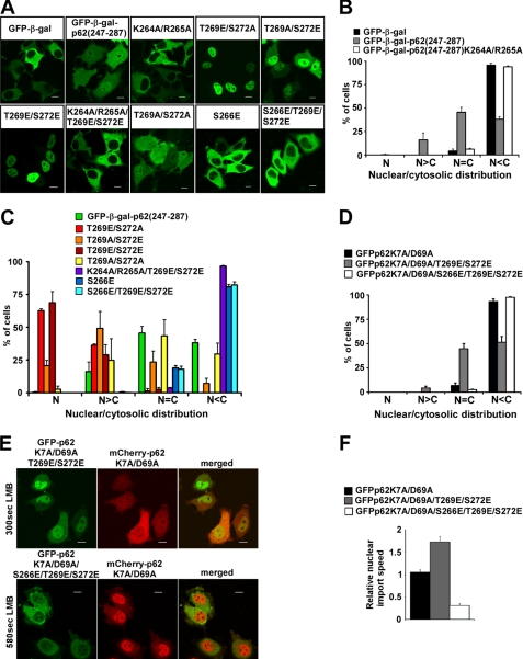
p62, also known as sequestosome1 (SQSTM1), A170, or ZIP, is a multifunctional protein implicated in several signal transduction pathways. p62 is induced by various forms of cellular stress, is degraded by autophagy, and acts as a cargo receptor for autophagic degradation of ubiquitinated targets. It is also suggested to shuttle ubiquitinated proteins for proteasomal degradation. p62 is commonly found in cytosolic protein inclusions in patients with protein aggregopathies, it is up-regulated in several forms of human tumors, and mutations in the gene are linked to classical adult onset Paget disease of the bone. To this end, p62 has generally been considered to be a cytosolic protein, and little attention has been paid to possible nuclear roles of this protein. Here, we present evidence that p62 shuttles continuously between nuclear and cytosolic compartments at a high rate. The protein is also found in nuclear promyelocytic leukemia bodies. We show that p62 contains two nuclear localization signals and a nuclear export signal. Our data suggest that the nucleocytoplasmic shuttling of p62 is modulated by phosphorylations at or near the most important nuclear localization signal, NLS2. The aggregation of p62 in cytosolic bodies also regulates the transport of p62 between the compartments. We found p62 to be essential for accumulation of polyubiquitinated proteins in promyelocytic leukemia bodies upon inhibition of nuclear protein export. Furthermore, p62 contributed to the assembly of proteasome-containing degradative compartments in the vicinity of nuclear aggregates containing polyglutamine-expanded Ataxin1Q84 and to the degradation of Ataxin1Q84.


Figures

FIGURE 1.
FIGURE 1.
p62 is a nucleocytoplasmic shuttling protein. A, endogenous p62 colocalizes with nuclear PML bodies in HeLa cells (upper panel) and translocates to the nucleus upon LMB treatment (lower panel). Scale bar, 10 μm. B and C, p62 is present in both nuclear and cytosolic compartments in prostate cancer tissue samples. The hematoxylin/eosin-stained slides of prostate cancer tissue microarrays were labeled with anti-p62 antibody and scored for the presence of p62 in the nucleus, cytosol, or both compartments. Numbers inside the bars represent the number of individual tissue samples assigned to each group. D, quantification of nuclear import speed of p62 is shown. HeLa cells were treated with LMB for indicated time periods, fixed, labeled with anti-p62 antibody, and scored as nuclear, nuclear/cytosolic, or cytosolic based on the intensity of p62 staining. Each time point represents the mean ± S.D. of three independent experiments with more than 200 cells counted per experiment.
FIGURE 2.
FIGURE 2.
The region of p62 between amino acids 303 and 320 is essential for the nuclear export of p62. A, the p62 region between amino acids 303 and 320 matches the consensus sequence of classical nuclear export signals. B, the region is recognized as an antigenic epitope by two commercially available mouse monoclonal antibodies. Shown is a Western blot (WB)of cell lysates from HeLa cells transiently transfected with the indicated constructs, developed with anti-p62 or anti-GFP antibodies (BD Transduction Laboratories #610833; Santa Cruz Biotechnology #28359). The bands corresponding to the GFP-p62 fusions and endogenous p62 are indicated as GFP-p62 and p62 endo., respectively. C–E, deletion of the region between amino acids 303–320 impairs the nuclear export of p62. C, shown are representative images of HeLa cells transiently transfected with the indicated DNA constructs or siRNA and stained with anti-p62 antibody (BD Transduction Laboratories #610833) where indicated. Scale bars equal 10 μm. D, immunoprecipitation (IP) of the indicated p62 constructs containing wild type or polymerization-impaired PB1 domain from cell lysates of transiently transfected HeLa cells is shown. E, quantification of nuclear/cytosolic distribution of transiently transfected p62 constructs from panel C is shown. N, only nuclear localization; N>C, nuclear staining is stronger than cytosolic; N=C, equal nucleocytosolic staining; N<C, cytosolic staining is stronger than nuclear. Each bar represents the mean ± S.D. of 3 independent experiments with more than 100 cells counted per experiment.
FIGURE 3.
FIGURE 3.
The region of p62 between amino acids 303 and 320 is sufficient for nuclear export. A, the nuclear export activity of p62 region 303–320 was tested using the pRev1.4-GFP assay system where putative NESs were inserted between the nuclear export-impaired Rev protein from HIV and GFP. B, representative images are shown of HeLa cells transiently transfected with Rev1.4-GFP, Rev1.4-NES-GFP, or Rev1.4-p62-(303–320)-GFP vectors (upper panel). Nucleocytoplasmic shuttling of Rev1.4-p62-(303–320)-GFP was verified by LMB treatment (lower panel). Scale bars equal 10 μm C, quantification of nuclear/cytosolic distribution of transiently transfected constructs from panel B is shown. Labels are the same as in Fig. 2E. Each bar represents the mean ± S.D. of 3 independent experiments with more than 100 cells counted per experiment. D, overexpression of the NES from Rev or the p62 fragment 303–320 inhibits the nuclear export of endogenous (end.) p62. Representative are shown of images of HeLa cells transiently transfected with indicated constructs, stained with anti-p62 polyclonal antibody. Scale bars equal 10 μm.
FIGURE 4.
FIGURE 4.
The region of p62 between amino acids 170 and 302 is sufficient for nuclear import. A, p62 contains two predicted basic nuclear localization sequences, NLS1, 186RKVK189 and NLS2, 264KRSR267. B and C, GFP fusion constructs of p62 fragment between amino acids 170 and 302 containing wild type (WT) or mutated NLS1 and NLS2 were transfected into HeLa cells. Twenty-four hours after transfection cells were scored based on the distribution of GFP fluorescence in the nuclear or cytosolic compartments. Representative images are shown in B. Scale bars equal 10 μm. Each bar in C represents the mean ± S.D. of 3 independent experiments with more than 100 cells counted per experiment.
FIGURE 5.
FIGURE 5.
NLS2 is essential for efficient nuclear import of p62. A, HeLa cells were transiently transfected with mCherry fusion construct of monomeric p62 (p62K7A/D69A; p62 shuttling reference control) together with the GFP fusion constructs of monomeric p62 containing wild type or mutated NLS1 and NLS2. Twenty-four hours after transfection, LMB was added to the culture medium to inhibit nuclear export of p62, and accumulation of GFP and mCherry fluorescence in the nuclei of the cells was imaged by live laser-scanning microscopy (B and C). The time needed for GFP- or mCherry-p62 fusion constructs to reach the same concentration in the nucleus as in the cytosol was measured and referred to as nuclear import time. To compensate for cell-to-cell variation, relative nuclear import time was calculated for each cell as a ratio between nuclear import time of GFP fusion constructs of wild type or NLS mutants (mut) of monomeric p62 and nuclear import time of the mCherry fusion construct of wild type, monomeric p62 (D). Each bar represents the mean relative nuclear import time ±S.D. for 20–35 cells from 3–6 independent experiments. Scale bars in panels B and C equal 10 μm.
FIGURE 6.
FIGURE 6.
Phosphomimetic mutations of Ser-266, Thr-269, and Ser-272 affect the efficiency of NLS2. The fragment of p62 encompassing amino acids 247 and 287 containing NLS2 was cloned in-frame with the EGFP-β-galactosidase (EGFP-gal) fusion protein. The resulting fusion construct was subjected to PCR mutagenesis to generate glutamate or alanine mutations of Ser-266, Thr-269, and Ser-272. Wild type or mutant variants of this construct were transiently transfected into HeLa cells (A), and their nuclear (N)/cytosolic (C) distribution was scored 24 h after transfection (B and C). Scale bars in panel A equal 10 μm. Each bar in panels B and C represents the mean ± S.D. of 3 independent experiments with more than 100 cells counted per experiment. D, constructs of full-length monomeric GFP-p62 with or without T269E/S272E or S266E/T269E/S272E mutations were transiently transfected into HeLa cells, and their nuclear/cytosolic distribution was scored 24 h after transfection. Each bar represents the mean ± S.D. of three independent experiments with more than 100 cells counted per experiment. E and F, GFP fusion constructs of full-length monomeric p62 with T269E/S272E or S266E/T269E/S272E were co-transfected with a mCherry fusion construct of full-length, monomeric p62. Twenty-four hours after transfection, cells were treated and imaged as described in the legend to Fig. 5. Cells with a similar nuclear/cytosolic distribution of GFP and mCherry fluorescence and with preferential cytosolic fluorescence before LMB treatment were selected for the calculation of nuclear import time. Relative nuclear import speed was calculated as a ratio between the nuclear import time of mCherry fusion construct of wild type monomeric p62 and the nuclear import times of GFP fusion constructs of phosphomimetic mutants of monomeric p62. Scale bars in panel E equal 10 μm. Each bar in panel F represents the mean relative nuclear import speed ± S.D. of more than 20 cells from 3–5 independent experiments.
FIGURE 7.
FIGURE 7.
p62 can recruit polyubiquitinated (polyUb) proteins to PML nuclear bodies. A, HeLa cells transfected with control or p62-targeting siRNA were left untreated or treated with LMB for 6 h, then fixed and stained with anti-polyubiquitin (FK1) and anti-p62 antibody. B, cells were transiently transfected with the pCW7-Myc-ubiquitin or pCW7-Myc-ubiquitin-K29R/K48R/K63R expression vectors and treated with LMB for 2 h at 24 h after transfection. The cells were fixed and stained with anti-Myc and anti-p62 antibodies. Scale bars equal 10 μm.
FIGURE 8.
FIGURE 8.
p62 is recruited to Ataxin1Q84 nuclear protein inclusions and facilitated the recruitment of proteasomes to these structures. A, quantification is shown of colocalization between nuclear GFP-Ataxin1Q84 inclusions and endogenous p62 in HeLa cells 24 and 48 h after transfection. HeLa cells were transiently transfected with the expression vector for GFP-Ataxin1Q84. 24 and 48 h after transfection cells were fixed, stained with antibodies against p62, and scored for colocalization between p62 and GFP-Ataxin1Q84. Each bar represents the mean ± S.D. of 3 independent experiments with more than 100 cells counted per experiment. B and C, confocal images are shown of HeLa cells transiently transfected with GFP-Ataxin1Q84, stained with antibodies against p62, PML, proteasome 20S core subunits, and polyubiquitin (polyUb) (FK1) 48 h after transfection. Scale bars equal 10 μm D, wild type (WT) or p62−/− MEFs were transiently transfected with the expression vector for GFP-Ataxin1Q84. 24 and 48 h after transfection, cells were fixed, stained with antibodies against the proteasome 20 S core subunits, and scored for colocalization between GFP-Ataxin1Q84 nuclear inclusions and proteasome staining by fluorescent microscopy. Each bar represents the mean ± S.D. of 3 independent experiments with more than 100 cells counted per experiment. E, HEK293 cells stably transfected with GFP-Ataxin1Q84 construct under the control of tet repressor-regulated CMV promoter were transfected with control (Ctrl.) siRNA or siRNA against p62, and the expression of GFP-Ataxin1Q84 was induced by tetracycline for 24 h. At 0, 12, 24, 48, and 72 h after removal of tetracycline, cells were lysed and subjected to SDS-PAGE and immunoblotting (WB) with the indicated antibodies. F, shown is quantification of the rate of GFP-Ataxin1Q84 degradation from E. The levels of GFP-Ataxin1Q84 were normalized to actin. Each bar represents the mean ± S.D. of three independent experiments. No ind., no induction.

Similar articles

Cited by

References

    1. Joung I., Strominger J. L., Shin J. (1996) Proc. Natl. Acad. Sci. U.S.A. 93, 5991–5995 - PMC - PubMed
    1. Puls A., Schmidt S., Grawe F., Stabel S. (1997) Proc. Natl. Acad. Sci. U.S.A. 94, 6191–6196 - PMC - PubMed
    1. Sanz L., Sanchez P., Lallena M. J., Diaz-Meco M. T., Moscat J. (1999) EMBO J. 18, 3044–3053 - PMC - PubMed
    1. Sanz L., Diaz-Meco M. T., Nakano H., Moscat J. (2000) EMBO J. 19, 1576–1586 - PMC - PubMed
    1. Wooten M. W., Seibenhener M. L., Neidigh K. B., Vandenplas M. L. (2000) Mol. Cell. Biol. 20, 4494–4504 - PMC - PubMed

Publication types

MeSH terms

LinkOut - more resources