

SSeCKS/Gravin/AKAP12 inhibits cancer cell invasiveness and chemotaxis by suppressing a protein kinase C- Raf/MEK/ERK pathway
- PMID: 20018890
- PMCID: PMC2836062
- DOI: 10.1074/jbc.M109.073494
SSeCKS/Gravin/AKAP12 inhibits cancer cell invasiveness and chemotaxis by suppressing a protein kinase C- Raf/MEK/ERK pathway
Abstract
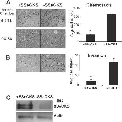
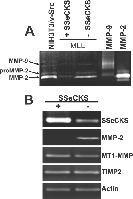
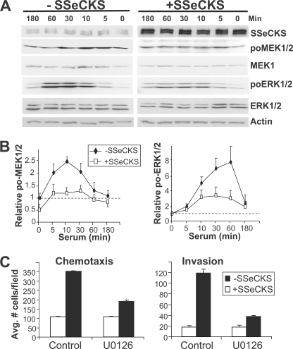
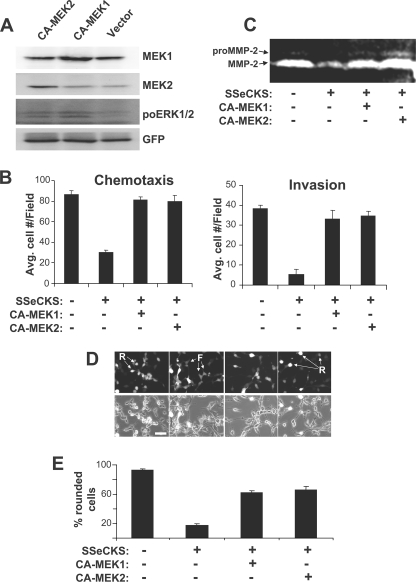
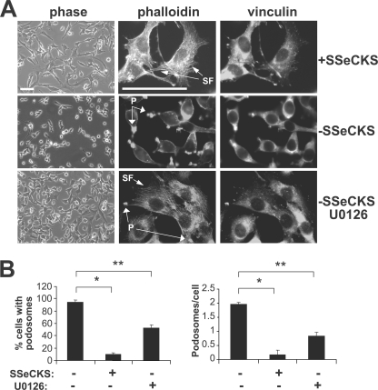
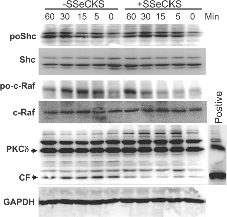
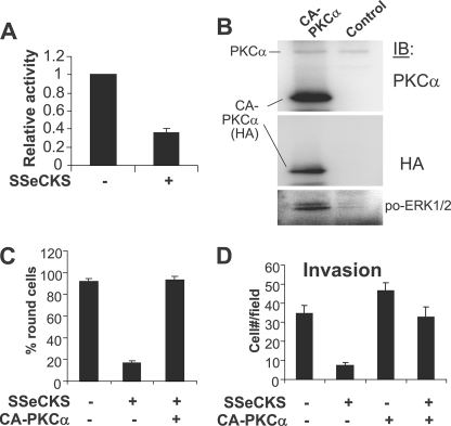
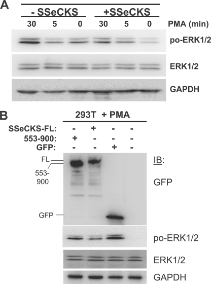
SSeCKS/Gravin/AKAP12 ("SSeCKS") encodes a cytoskeletal protein that regulates G(1) --> S progression by scaffolding cyclins, protein kinase C (PKC) and PKA. SSeCKS is down-regulated in many tumor types including prostate, and when re-expressed in MAT-LyLu (MLL) prostate cancer cells, SSeCKS selectively inhibits metastasis by suppressing neovascularization at distal sites, correlating with its ability to down-regulate proangiogenic genes including Vegfa. However, the forced re-expression of VEGF only rescues partial lung metastasis formation. Here, we show that SSeCKS potently inhibits chemotaxis and Matrigel invasion, motility parameters contributing to metastasis formation. SSeCKS suppressed serum-induced activation of the Raf/MEK/ERK pathway, resulting in down-regulation of matrix metalloproteinase-2 expression. In contrast, SSeCKS had no effect on serum-induced phosphorylation of the Src substrate, Shc, in agreement with our previous data that SSeCKS does not inhibit Src kinase activity in cells. Invasiveness and chemotaxis could be restored by the forced expression of constitutively active MEK1, MEK2, ERK1, or PKCalpha. SSeCKS suppressed phorbol ester-induced ERK1/2 activity only if it encoded its PKC binding domain (amino acids 553-900), suggesting that SSeCKS attenuates ERK activation through a direct scaffolding of conventional and/or novel PKC isozymes. Finally, control of MLL invasiveness by SSeCKS is influenced by the actin cytoskeleton: the ability of SSeCKS to inhibit podosome formation is unaffected by cytochalasin D or jasplakinolide, whereas its ability to inhibit MEK1/2 and ERK1/2 activation is nullified by jasplakinolide. Our findings suggest that SSeCKS suppresses metastatic motility by disengaging activated Src and then inhibiting the PKC-Raf/MEK/ERK pathways controlling matrix metalloproteinase-2 expression and podosome formation.
Figures
Similar articles
-
Suppression of tumor and metastasis progression through the scaffolding functions of SSeCKS/Gravin/AKAP12.Cancer Metastasis Rev. 2012 Dec;31(3-4):493-500. doi: 10.1007/s10555-012-9360-1. Cancer Metastasis Rev. 2012. PMID: 22684366 Free PMC article. Review.
-
Control of protein kinase C activity, phorbol ester-induced cytoskeletal remodeling, and cell survival signals by the scaffolding protein SSeCKS/GRAVIN/AKAP12.J Biol Chem. 2011 Nov 4;286(44):38356-38366. doi: 10.1074/jbc.M111.258830. Epub 2011 Sep 7. J Biol Chem. 2011. PMID: 21903576 Free PMC article.
-
Adhesion-mediated cytoskeletal remodeling is controlled by the direct scaffolding of Src from FAK complexes to lipid rafts by SSeCKS/AKAP12.Oncogene. 2013 Apr 18;32(16):2016-26. doi: 10.1038/onc.2012.218. Epub 2012 Jun 18. Oncogene. 2013. PMID: 22710722 Free PMC article.
-
SSeCKS/Gravin/AKAP12 metastasis suppressor inhibits podosome formation via RhoA- and Cdc42-dependent pathways.Mol Cancer Res. 2006 Mar;4(3):151-8. doi: 10.1158/1541-7786.MCR-05-0252. Mol Cancer Res. 2006. PMID: 16547152
-
The role of SSeCKS/gravin/AKAP12 scaffolding proteins in the spaciotemporal control of signaling pathways in oncogenesis and development.Front Biosci. 2002 Aug 1;7:d1782-97. doi: 10.2741/A879. Front Biosci. 2002. PMID: 12133808 Review.
Cited by
-
EphA6 promotes angiogenesis and prostate cancer metastasis and is associated with human prostate cancer progression.Oncotarget. 2015 Sep 8;6(26):22587-97. doi: 10.18632/oncotarget.4088. Oncotarget. 2015. PMID: 26041887 Free PMC article.
-
Suppression of tumor and metastasis progression through the scaffolding functions of SSeCKS/Gravin/AKAP12.Cancer Metastasis Rev. 2012 Dec;31(3-4):493-500. doi: 10.1007/s10555-012-9360-1. Cancer Metastasis Rev. 2012. PMID: 22684366 Free PMC article. Review.
-
The short inverted repeats-induced circEXOC6B inhibits prostate cancer metastasis by enhancing the binding of RBMS1 and HuR.Mol Ther. 2023 Jun 7;31(6):1705-1721. doi: 10.1016/j.ymthe.2022.08.006. Epub 2022 Aug 15. Mol Ther. 2023. PMID: 35974702 Free PMC article.
-
Metastasis suppressor genes at the interface between the environment and tumor cell growth.Int Rev Cell Mol Biol. 2011;286:107-80. doi: 10.1016/B978-0-12-385859-7.00003-3. Int Rev Cell Mol Biol. 2011. PMID: 21199781 Free PMC article. Review.
-
Role of the polypeptide N-acetylgalactosaminyltransferase 3 in ovarian cancer progression: possible implications in abnormal mucin O-glycosylation.Oncotarget. 2014 Jan 30;5(2):544-60. doi: 10.18632/oncotarget.1652. Oncotarget. 2014. PMID: 24504219 Free PMC article.
References
-
- Lin X., Tombler E., Nelson P. J., Ross M., Gelman I. H. (1996) J. Biol. Chem. 271, 28430–28438 - PubMed
-
- Chapline C., Cottom J., Tobin H., Hulmes J., Crabb J., Jaken S. (1998) J. Biol. Chem. 273, 19482–19489 - PubMed
-
- Gelman I. H., Lee K., Tombler E., Gordon R., Lin X. (1998) Cell Motil. Cytoskeleton 41, 1–17 - PubMed
-
- Nelson P. J., Moissoglu K., Vargas J., Jr., Klotman P. E., Gelman I. H. (1999) J. Cell Sci. 112, 361–370 - PubMed
Publication types
MeSH terms
Substances
Grants and funding
LinkOut - more resources
Full Text Sources
Research Materials
Miscellaneous
