

ATP hydrolysis in Eg5 kinesin involves a catalytic two-water mechanism
- PMID: 20018897
- PMCID: PMC2820811
- DOI: 10.1074/jbc.M109.071233
ATP hydrolysis in Eg5 kinesin involves a catalytic two-water mechanism
Abstract
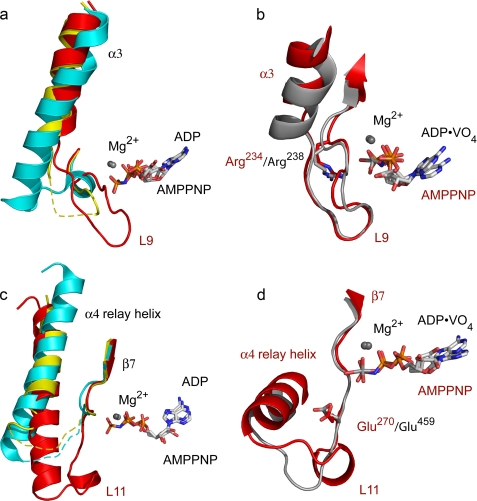
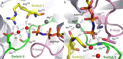
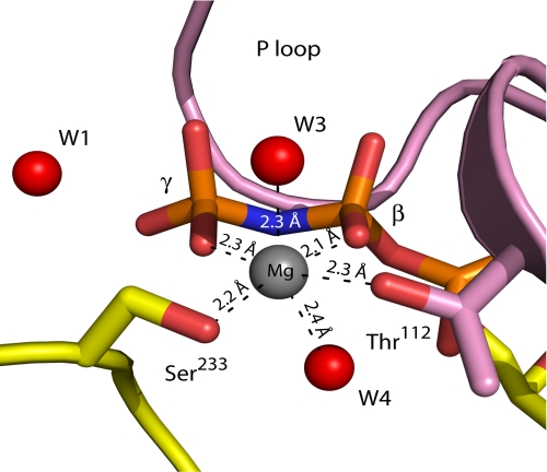
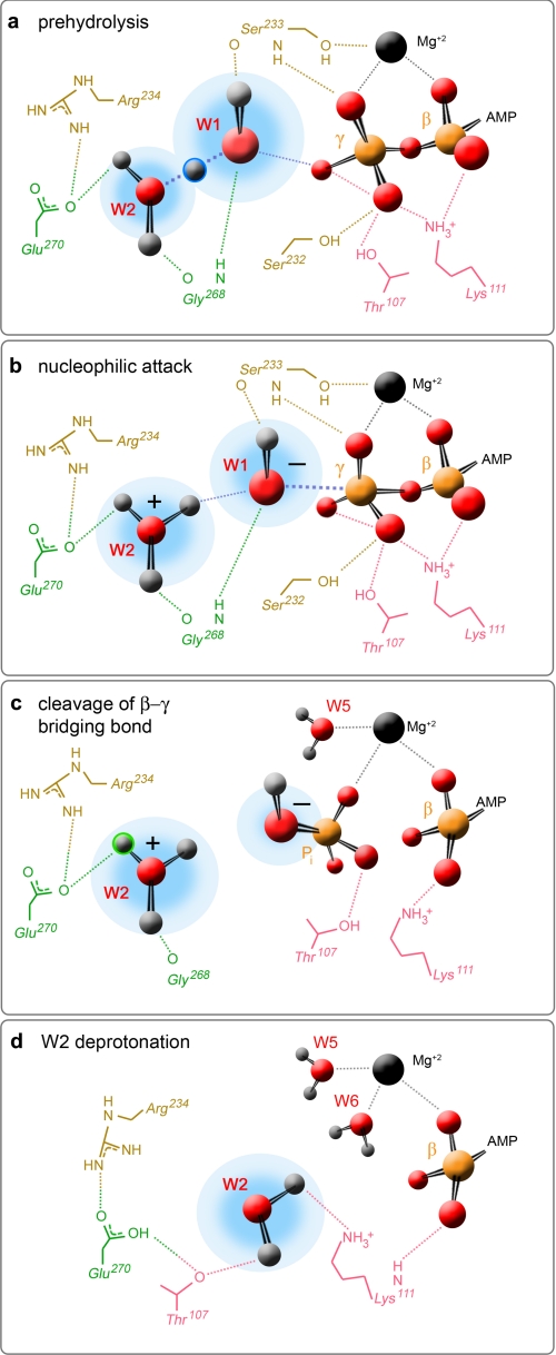
Motor proteins couple steps in ATP binding and hydrolysis to conformational switching both in and remote from the active site. In our kinesin.AMPPPNP crystal structure, closure of the active site results in structural transformations appropriate for microtubule binding and organizes an orthosteric two-water cluster. We conclude that a proton is shared between the lytic water, positioned for gamma-phosphate attack, and a second water that serves as a general base. To our knowledge, this is the first experimental detection of the catalytic base for any ATPase. Deprotonation of the second water by switch residues likely triggers subsequent large scale structural rearrangements. Therefore, the catalytic base is responsible for initiating nucleophilic attack of ATP and for relaying the positive charge over long distances to initiate mechanotransduction. Coordination of switch movements via sequential proton transfer along paired water clusters may be universal for nucleotide triphosphatases with conserved active sites, such as myosins and G-proteins.
Figures

Similar articles
-
Real-time structural transitions are coupled to chemical steps in ATP hydrolysis by Eg5 kinesin.J Biol Chem. 2010 Apr 9;285(15):11073-7. doi: 10.1074/jbc.C110.103762. Epub 2010 Feb 12. J Biol Chem. 2010. PMID: 20154092 Free PMC article.
-
Structural snapshots of the kinesin-2 OSM-3 along its nucleotide cycle: implications for the ATP hydrolysis mechanism.FEBS Open Bio. 2021 Mar;11(3):564-577. doi: 10.1002/2211-5463.13101. Epub 2021 Feb 28. FEBS Open Bio. 2021. PMID: 33513284 Free PMC article.
-
Large conformational changes in a kinesin motor catalyzed by interaction with microtubules.Mol Cell. 2006 Sep 15;23(6):913-23. doi: 10.1016/j.molcel.2006.07.020. Mol Cell. 2006. PMID: 16973442 Free PMC article.
-
Review: Mechanochemistry of the kinesin-1 ATPase.Biopolymers. 2016 Aug;105(8):476-82. doi: 10.1002/bip.22862. Biopolymers. 2016. PMID: 27120111 Free PMC article. Review.
-
Kinesin: switch I & II and the motor mechanism.J Cell Sci. 2002 Jan 1;115(Pt 1):15-23. doi: 10.1242/jcs.115.1.15. J Cell Sci. 2002. PMID: 11801720 Review.
Cited by
-
No evidence of locus heterogeneity in familial microcephaly with or without chorioretinopathy, lymphedema, or mental retardation syndrome.Orphanet J Rare Dis. 2015 May 2;10:52. doi: 10.1186/s13023-015-0271-4. Orphanet J Rare Dis. 2015. PMID: 25934493 Free PMC article.
-
The loop 5 element structurally and kinetically coordinates dimers of the human kinesin-5, Eg5.Biophys J. 2011 Dec 7;101(11):2760-9. doi: 10.1016/j.bpj.2011.10.032. Biophys J. 2011. PMID: 22261065 Free PMC article.
-
Microtubule capture by mitotic kinesin centromere protein E (CENP-E).J Biol Chem. 2012 Jul 20;287(30):24894-904. doi: 10.1074/jbc.M112.376830. Epub 2012 May 27. J Biol Chem. 2012. PMID: 22637578 Free PMC article.
-
Analysis of Biphenyl-Type Inhibitors Targeting the Eg5 α4/α6 Allosteric Pocket.ACS Omega. 2017 May 31;2(5):1836-1849. doi: 10.1021/acsomega.6b00467. Epub 2017 May 5. ACS Omega. 2017. PMID: 30023646 Free PMC article.
-
Kinesin-2 KIF3AC and KIF3AB Can Drive Long-Range Transport along Microtubules.Biophys J. 2015 Oct 6;109(7):1472-82. doi: 10.1016/j.bpj.2015.08.004. Biophys J. 2015. PMID: 26445448 Free PMC article.
References
-
- Endow S. A. (1999) Eur. J. Biochem. 262, 12–18 - PubMed
-
- Hirokawa N. (1998) Science 279, 519–526 - PubMed
-
- Fisher A. J., Smith C. A., Thoden J. B., Smith R., Sutoh K., Holden H. M., Rayment I. (1995) Biochemistry 34, 8960–8972 - PubMed
-
- Gulick A. M., Bauer C. B., Thoden J. B., Rayment I. (1997) Biochemistry 36, 11619–11628 - PubMed
-
- Onishi H., Mochizuki N., Morales M. F. (2004) Biochemistry 43, 3757–3763 - PubMed
Publication types
MeSH terms
Substances
Associated data
- Actions
Grants and funding
LinkOut - more resources
Full Text Sources
Other Literature Sources
