. 2010 Feb 12;285(7):4366-74.
doi: 10.1074/jbc.M109.066092. Epub 2009 Dec 15.

Annexin A2 is a molecular target for TM601, a peptide with tumor-targeting and anti-angiogenic effects

Affiliations

Annexin A2 is a molecular target for TM601, a peptide with tumor-targeting and anti-angiogenic effects

Kamala Kesavan et al. J Biol Chem. .

Abstract

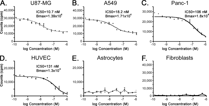
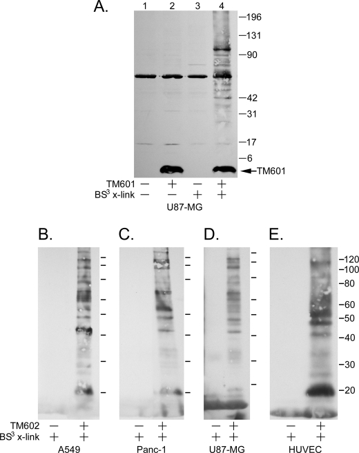
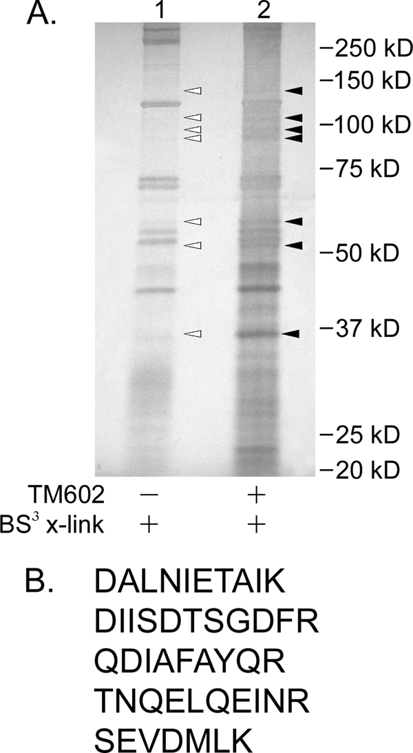
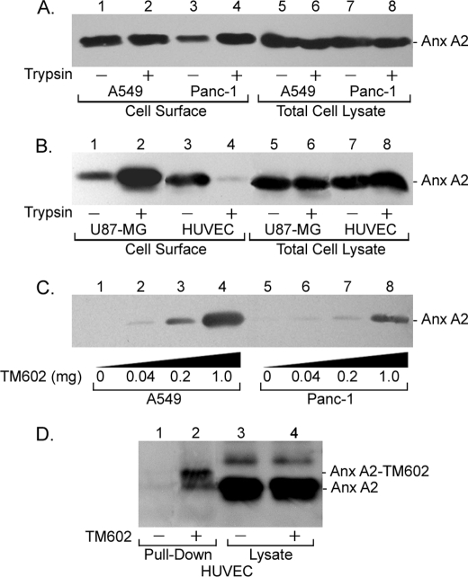
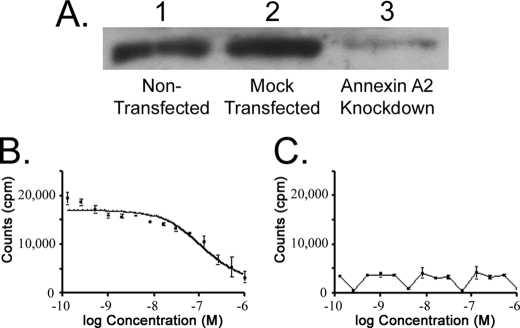
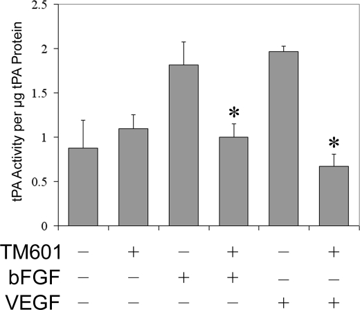
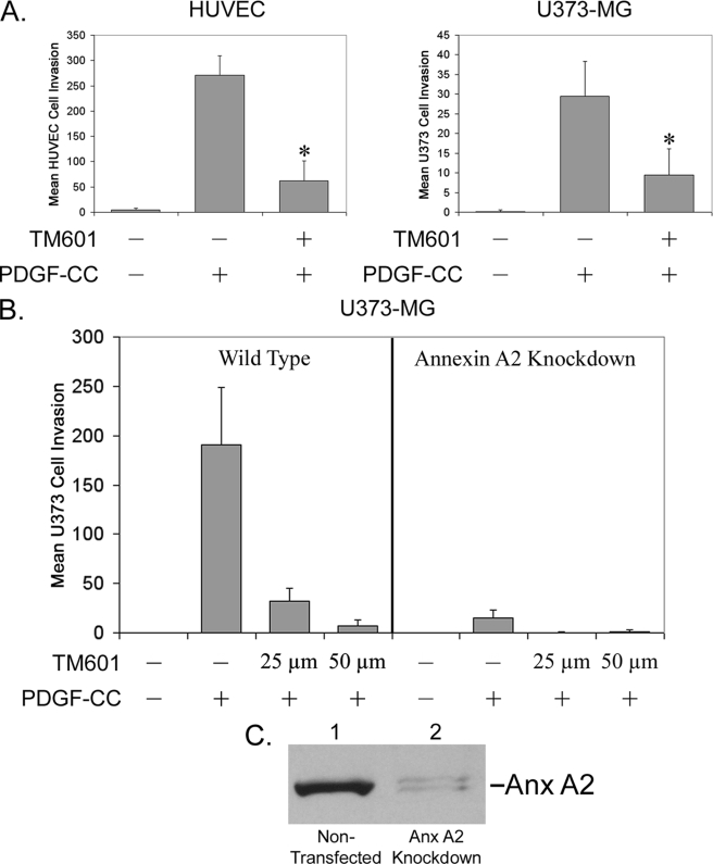
TM601 is a synthetic form of chlorotoxin, a 36-amino acid peptide derived from the venom of the Israeli scorpion, Leirius quinquestriatus, initially found to specifically bind and inhibit the migration of glioma cells in culture. Subsequent studies demonstrated specific in vitro binding to additional tumor cell lines. Recently, we demonstrated that proliferating human vascular endothelial cells are the only normal cell line tested that exhibits specific binding to TM601. Here, we identify annexin A2 as a novel binding partner for TM601 in multiple human tumor cell lines and human umbilical vein endothelial cell (HUVEC). We demonstrate that the surface binding of TM601 to the pancreatic tumor cell line Panc-1 is dependent on the expression of annexin A2. Identification of annexin A2 as a binding partner for TM601 is also consistent with the anti-angiogenic effects of TM601. Annexin A2 functions in angiogenesis by binding to tissue plasminogen activator and regulating plasminogen activation on vascular endothelial cells. We demonstrate that in HUVECs, TM601 inhibits both vascular endothelial growth factor- and basic fibroblast growth factor-induced tissue plasminogen activator activation, which is required for activation of plasminogen to plasmin. Consistent with inhibition of cell surface protease activity, TM601 also inhibits platelet-derived growth factor-C induced trans-well migration of both HUVEC and U373-MG glioma cells.


Figures

FIGURE 1.
FIGURE 1.
Specific binding of TM601 to the cell surface of tumor and vascular endothelial cells. Cell surface binding of TM601 was quantified by measuring the amount of technetium-99m-labeled TM601 bound to the cell surface at 4 °C in the presence of unlabeled TM601 monomer used as competitor. TM601 binds the surface of multiple tumor cells with a high affinity for U87-MG glioma (A) and A549 lung carcinoma (B) and lower affinity for Panc-1 pancreatic carcinoma (C) cell lines. TM601 binds HUVECs (D) in culture with an affinity comparable with Panc-1 cells and shows little surface binding to either normal human astrocytes (E) or normal human dermal fibroblasts (F).
FIGURE 2.
FIGURE 2.
TM601 associates with a specific subset of cell surface proteins in both tumor and HUVECs. A, lysates from treated and untreated (±TM601 at 4 °C) U87-MG glioma cells chemically cross-linked with a membrane-impermeable cross-linking agent (BS3) were separated by SDS-PAGE and immunoblotted with anti-TM601 antibodies (A). Comparison of lane 2 (TM601 without cross-linking) and lane 3 (BS3 cross-linking without TM601) with lane 4 (TM601 followed by BS3 cross-linking) reveals a specific subset of cross-linked gel-shifted cell surface proteins that associate with TM601. B–E, the associating proteins can be isolated using TM602 (biotinylated TM601) in a pulldown assay with neutrAvidinTM beads. Anti-TM601 Western blot of neutrAvidinTM pulldowns from lysates of TM602 treated (+) and untreated cells (−) chemically cross-linked with BS3 shows a similar subset of surface proteins specifically associating with TM602 on A549 (B), Panc-1 (C), and U87-MG (D) tumor cell lines and HUVECs (E).
FIGURE 3.
FIGURE 3.
Mass spectrometric identification of TM602 binding partner expressed on tumor cell surface. A, lysates from treated and untreated (±TM602 at 4 °C) U87-MG glioma cells chemically cross-linked with membrane-impermeable cross-linking agent (BS3) and isolated by neutrAvidinTM pulldown were separated by SDS-PAGE and silver-stained using the SilverQuestTM staining kit. The indicated bands were extracted from control (lane 1, open triangles) and TM602 pulldown samples (lane 2, filled triangles) and analyzed by mass spectrometry to identify isolated protein sequences. As indicated, comparisons were made between mass spectrometric results of gel slices of identical molecular mass (kDa) between the two lanes to identify proteins specifically enriched in TM602-containing lane. B, peptides identified exclusively in lane 2 that have sequence identity to human annexin A2.
FIGURE 4.
FIGURE 4.
TM602 binds annexin A2 expressed on the surface of multiple tumor and vascular endothelial cells. Confluent monolayers of A549, Panc-1, U87-MG, and HUVECs treated with or without trypsin were surface-biotinylated, and biotinylated proteins were isolated using neutrAvidinTM beads. The levels of annexin A2 expressed on the surface of tumor cell lines and HUVECs and the sensitivity to trypsin were determined using an anti-annexin A2 Western blot. A, annexin A2 levels at the cell surface and in total cell lysates for plate-bound (lanes 1, 3, 5, and 7) and trypsinized (lanes 2, 4, 6, and 8) A549 (lanes 1, 2, 5, and 6) and Panc-1 (lanes 3, 4, 7, and 8) cells. B, annexin A2 levels at the cell surface and in total cell lysates for plate-bound (lanes 1, 3, 5, and 7) and trypsinized (lanes 2, 4, 6, and 8) U87-MG (lanes 1, 2, 5, and 6) glioma and HUVECs (lanes 3, 4, 7, and 8). Annexin A2 is expressed at the cell surface in all the aforementioned cell lines. In HUVECs, surface annexin A2 is sensitive to trypsinization (B, lanes 3 and 4). C and D, the association of TM602 with cell surface annexin A2 was validated using an anti-annexin A2 Western blot of neutrAvidinTM pulldowns of lysates from A549, Panc-1, and HUVECs following TM602 surface cross-linking. Annexin A2 specifically associates with TM602 at the cell surface of A549 and Panc-1 tumor cells (C) as well as HUVECs (D).
FIGURE 5.
FIGURE 5.
Annexin A2 mediates cell surface binding of TM601 to Panc-1 pancreatic carcinoma cell line. Functional importance of the interaction between annexin A2 and TM601 was tested using a siRNA knockdown approach in Panc-1 carcinoma cells. A, Western blot using anti-annexin A2 antibody shows that the level of annexin A2 in untransfected Panc-1 cells was comparable with that of mock-transfected cells (lanes 1 and 2). Annexin A2 siRNA transfected cells showed a significant reduction of annexin A2 levels at 48 h post-transfection by annexin A2 Western blot of total cell lysates. As shown in A, the level of annexin A2 is similar in untransfected and mock-transfected cells (lanes 1 and 2) and is significantly decreased in annexin A2 siRNA treated cells (lane 3). Control and annexin A2 siRNA-treated Panc-1 cells were also tested for surface 99mTc-TM601 binding assays. B, competitive binding of 99mTc-TM601 in untransfected Panc-1 cells. C, competitive binding of 99mTc-TM601 to surface of Panc-1 cells transfected with annexin A2 siRNA. Annexin A2 siRNA-mediated knockdown in Panc-1 cells abolished the surface binding of TM601.
FIGURE 6.
FIGURE 6.
TM601 inhibits VEGF-stimulated tPA activity in human vascular endothelial cell supernatants. The effect of TM601 on VEGF- and bFGF-stimulated activation of secreted tPA in HUVECs was measured. Both VEGF- and bFGF-stimulated tPA activity in serum-starved HUVECs and TM601 treatment significantly decreased the amount of active tPA present in the supernatants of vascular endothelial cells treated with either VEGF or bFGF. *, p < 0.05. Bar, S.D.
FIGURE 7.
FIGURE 7.
TM601 inhibits PDGF-CC-induced U373-MG glioma and HUVEC migration in trans-well chamber assays. A, in a trans-well assay using HUVEC or U373-MG cells, PDGF-CC strongly stimulates cell migration, and TM601 significantly inhibits the PDGF-CC-stimulated migration of both vascular endothelial cells and U373-MG glioma cells. *, p < 0.05. Bar, S.D. B, functional importance of annexin A2 in PDGF-CC-stimulated glioma migration was tested using an annexin A2 siRNA knockdown approach. Mock-transfected wild-type U373-MG and annexin A2 knockdown U373-MG glioma cells were used in a trans-well assay testing migration stimulated by PDGF-CC. Wild-type cells demonstrated significant migration stimulated by PDGF-CC that was inhibited by TM601 treatment. Annexin A2 knockdown cells demonstrated very weak migration stimulated by PDGF-CC that was completely abolished by a lower dose of TM601 (25 μm) as compared with wild-type cells. C, Western blot using anti-annexin A2 antibody demonstrates the levels of annexin A2 after annexin A2 siRNA treatment.

Similar articles

Cited by

References

    1. DeBin J. A., Maggio J. E., Strichartz G. R. (1993) Am. J. Physiol. 264, C361–C369 - PubMed
    1. Mamelak A. N., Jacoby D. B. (2007) Expert Opin. Drug Deliv. 4, 175–186 - PubMed
    1. Mamelak A. N., Rosenfeld S., Bucholz R., Raubitschek A., Nabors L. B., Fiveash J. B., Shen S., Khazaeli M. B., Colcher D., Liu A., Osman M., Guthrie B., Schade-Bijur S., Hablitz D. M., Alvarez V. L., Gonda M. A. (2006) J. Clin. Oncol. 24, 3644–3650 - PubMed
    1. Gibbin T. E., Senzer N., Raizer J. J., Shen J., Nabors L. B., Wiranowska M., Fiveash J. B. (2009) J. Clin. Oncol. 27, e14507
    1. Ullrich N., Gillespie G. Y., Sontheimer H. (1995) Neuroreport 7, 343–347 - PubMed

LinkOut - more resources