. 2010 Mar;298(3):C496-509.
doi: 10.1152/ajpcell.00464.2009. Epub 2009 Dec 16.

Tetramerization domain mutations in KCNA5 affect channel kinetics and cause abnormal trafficking patterns

Affiliations

Tetramerization domain mutations in KCNA5 affect channel kinetics and cause abnormal trafficking patterns

Elyssa D Burg et al. Am J Physiol Cell Physiol. 2010 Mar.

Abstract

The activity of voltage-gated K(+) (K(V)) channels plays an important role in regulating pulmonary artery smooth muscle cell (PASMC) contraction, proliferation, and apoptosis. The highly conserved NH(2)-terminal tetramerization domain (T1) of K(V) channels is important for proper channel assembly, association with regulatory K(V) beta-subunits, and localization of the channel to the plasma membrane. We recently reported two nonsynonymous mutations (G182R and E211D) in the KCNA5 gene of patients with idiopathic pulmonary arterial hypertension, which localize to the T1 domain of KCNA5. To study the electrophysiological properties and expression patterns of the mutants compared with the wild-type (WT) channel in vitro, we transfected HEK-293 cells with WT KCNA5, G182R, E211D, or the double mutant G182R/E211D channel. The mutants form functional channels; however, whole cell current kinetic differences between WT and mutant channels exist. Steady-state inactivation curves of the G182R and G182R/E211D channels reveal accelerated inactivation; the mutant channels inactivated at more hyperpolarized potentials compared with the WT channel. Channel protein expression was also decreased by the mutations. Compared with the WT channel, which was present in its mature glycosylated form, the mutant channels are present in greater proportion in their immature form in HEK-293 cells. Furthermore, G182R protein level is greatly reduced in COS-1 cells compared with WT. Immunostaining data support the hypothesis that, while WT protein localizes to the plasma membrane, mutant protein is mainly retained in intracellular packets. Overall, these data support a role for the T1 domain in channel kinetics as well as in KCNA5 channel subcellular localization.


Figures

Fig. 1.
Fig. 1.
Two nonsynonymous mutations identified in the KCNA5 gene from idiopathic pulmonary arterial hypertension (IPAH) patients localize to the NH2-terminal tetramerization domain (T1 domain). A, left: schematic diagram of voltage-gated K+ (KV) channel subunit showing 6 transmembrane domains (S1–S6), cytosolic NH2 and COOH termini, and the K+ selective pore region (P). Right: the T1 domain mediates both KCNA (the pore-forming α-subunit) assembly and KCNA-KCNAB (the regulatory β-subunit, KVβ) interactions to form tetrameric channels. B: chromatograms of the regions containing G182R (middle) and E211D (bottom) (boxed in red) are shown. Sequence from normal subject is shown as control (top). C: alignment of the T1 region encompassing the G182 and E211 residues from various human KCNA, KCNB, KCNC, and KCND channels. Residues that correspond to G182 or E211 are shown in bold. Sequences are from the NCBI database with the following accession numbers: KCNA1: NM_000217; KCNA2: NM_004974; KCNA3: NM_002232; KCNA4: NM_002233; KCNA5: NM_002234; KCNA6: NM_002235; KCNA7: NM_031886; KCNA10: NM_005549; KCNB1: NM_004975; KCNB2: NM_004770; KCNC1: NM_004976; KCNC2: NM_139136; KCNC3: NM_004977; KCNC4: NM_004978; KCND1: NM_004979; KCND2: NM_012281; KCND3: NM_004980. D, a: ribbon diagram of wild-type (WT) KCNA5 with side chains of the glycine at position 182 (G182) and the glutamate at position 211 (E211) indicated. b: Space filling diagram of WT KCNA5 with the side chains of glycine (G182) and glutamate (E211) indicated. c: Space filling diagram of mutant KCNA5 (c) with the side chains of the arginine at position 182 (R182) and the aspartate at position 211 (D211) indicated. Four differently colored subunits are shown, but the residues are indicated only on the orange subunit for clarity. The top of the channel is extracellular, while the bottom is cytoplasmically located. Close-ups of the region of interest are shown at bottom.
Fig. 2.
Fig. 2.
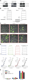
Mutant KCNA5 forms functional homotetrameric channels. A: COS-1, HEK-293, and human pulmonary artery smooth muscle cells (PASMC) were transiently transfected with either empty vector [green fluorescent protein (GFP)] or WT KCNA5 (WT) as indicated. Whole cell lysate was probed on immunoblot for KCNA5 (67 kDa) and GAPDH as a loading control. B: HEK or COS cells were transiently transfected with water (Mock) or WT KCNA5 (+KCNA5) and subjected to a standard current-voltage (I-V) pulse protocol. The pulse protocol stepped from −80 to +60 mV in 20-mV increments from a holding potential of −70 mV (inset). Representative recordings of whole cell KV currents [IK(V)] are shown. C: HEK cells were transiently transfected with water (Mock), empty vector [enhanced GFP (EGFP)], WT KCNA5, G182R, E211D, or G182R/E211D. Transfected cells were identified by green fluorescence and used for patch-clamp experiments. D: representative recordings of IK(V) from HEK cells transfected with the indicated vectors and constructs. Representative IK(V) (top) and summarized I-V curves (bottom) from the cells transfected with WT KCNA5 (n = 15), G182R (n = 15), E211D (n = 14), or G182R/E211D (n = 16 cells) are shown. E: summarized amplitudes of IK(V) at −60, −40, and −20 mV indicate that the activation threshold of the currents is close to −40 mV. All data are presented as means ± SE.
Fig. 3.
Fig. 3.
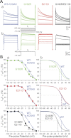
Voltage-dependent inactivation is accelerated in the G182R mutant KCNA5 channel. A standard 2-pulse inactivation protocol was used to determine channel availability after a 10-s prepulse in HEK-293 cells transiently transfected with WT KCNA5, G182R, E211D, or G182R/E211D. A: representative recordings of currents, elicited by prepulse (t1) and test pulse (t2) from cells transfected with the indicated vector are shown in a; the recordings of the currents elicited by t2 are enlarged and shown in b. B: inactivation curves (a) are plotted as current amplitude during test pulse (It2) over the amplitude during prepulse (It1) against the prepulse potential (It2/It1). The Boltzmann equation was used to fit the inactivation curves and determine V1/2 (b). All data are presented as means ± SE. WT KCNA5, n = 12; G182R, n = 12; E211D, n = 12; G182R/E211D, n = 14. *P < 0.05, ***P < 0.001 vs. WT KCNA5.
Fig. 4.
Fig. 4.
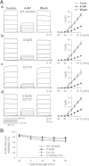
Mutations in KCNA5 at G182 and E211 do not affect the pharmacological effect of 4-aminopyridine (4-AP). A: a standard I-V pulse protocol was delivered to HEK-293 cells transiently transfected with the indicated vector [WT KCNA5 (a), G182R (b), E211D (c), or G182R/E211D (d)]. Currents were first recorded in standard physiological salt solution (Control), then with the addition to the bathing solution of 5 mM 4-AP, and finally after washout of 4-AP in physiological salt solution (Wash). I-V curves are shown on right. B: summarized data are shown as % inhibition of IK(V) during 4-AP exposure at different test potentials (−20, 0, 20, 40, and 60 mV). WT KCNA5, n = 15; G182R, n = 8; E211D, n = 8; G182R/E211D, n = 8. Data are presented as means ± SE. Data were compared with ANOVA and Tukey's post hoc test; no significant differences were found.
Fig. 5.
Fig. 5.
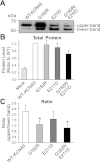
G182R and E211D mutations cause incomplete processing of KCNA5 in HEK-293 cells. HEK-293 cells were transfected with WT KCNA5, G182R, E211D, or G182R/E211D and subjected to standard immunoblot procedures. A: representative immunoblot from cells transfected with the indicated constructs. Blots were probed for KCNA5 (67 kDa). B: summarized data of KCNA5 protein levels in HEK-293 cells transfected with water (Mock), G182R, E211D, or G182R/E211D are presented as ratio to WT KCNA5 levels (n = 13). C: summarized data of upper- to lower-band ratios are shown (n = 7). Data are presented as means ± SE. Data were compared with ANOVA and Tukey's post hoc analysis. *P < 0.05 compared with WT-KCNA5.
Fig. 6.
Fig. 6.
Mutant KCNA5 is located in perinuclear packets and not on the cell surface of transfected HEK-293 cells and human (h)PASMC. A: HEK-293 cells transfected with WT KCNA5 (a and c) or G182R (b and d) were stained with anti-KCNA5 antibody (Ab-KCNA5, red) and 4′,6′-diamidino-2-phenylindole (DAPI, blue). Transfected cells were identified by the presence of EGFP (green). Images of cells transfected with WT KCNA5 or G182R and stained for KCNA5 are enlarged for clarity (c and d). B: mock-transfected HEK-293 cells exposed to antibody (Ab-KCNA5) demonstrate anti-KCNA5 antibody specificity (a), and WT-KCNA5-transfected cells without antibody treatment [(−)Ab-KCNA5] demonstrate secondary antibody specificity (b). C: hPASMC transfected with WT KCNA5 (a) or G182R/E211D (b) were stained as in A. All fields are shown at ×60 magnification except Ac and Ad, which are digitally enlarged images of Aa and Ab, respectively.
Fig. 7.
Fig. 7.
G182R protein expression is significantly decreased in COS-1 cells. A: COS-1 cells were transiently transfected with water (Mock), WT-KCNA5, G182R, E211D, or G182R/E211D. Representative images are shown at ×40 magnification. B: transfected cells were subjected to standard immunoblot procedure to detect KCNA5 (67 kDa) or GAPDH (36 kDa) proteins. Summarized data are expressed in arbitrary units as the band intensity of KCNA5 divided by that of GAPDH (n = 4). C: transfected COS-1 cells were subjected to RT-PCR to detect KCNA5 or GAPDH mRNA. Summarized data are expressed as arbitrary units of KCNA5 normalized to GAPDH (n = 2). Data are presented as means ± SE and were compared with ANOVA and Tukey's post hoc analysis. *P < 0.05, ***P < 0.001 compared with WT KCNA5.
Fig. 8.
Fig. 8.
Cotransfection of KVβ subunits affects KCNA5 channel kinetics. HEK-293 cells were transfected with WT KCNA5 alone (KCNA5) or in the presence of KVβ1.3-hemagglutinin (HA) (KCNA5/KVβ1.3). A and B: representative current recordings (A) and I-V curves (B) are shown (pulse protocol, bottom). C: averaged currents (a) and normalized currents (I/Imax, b) at +60 mV in cells transiently transfected with WT KCNA5 alone (KCNA5) or WT KCNA5 + KVβ1.3-HA (KCNA5+KVβ1.3).
Fig. 9.
Fig. 9.
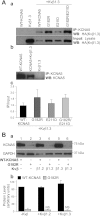
Decreased G182R expression in COS-1 cells cannot be rescued by overexpression of KVβ subunits. A, a: HEK-293 cells were transfected with WT KCNA5 or KVβ1.3-HA alone or cotransfected with WT KCNA5, G182R, E211D, or G182R/E211D and KVβ1.3-HA. Whole cell protein lysate was immunoprecipitated (IP) with anti-KCNA5 antibody and immunoblotted for HA tag [IP: KCNA5, Western blot (WB): HA; top]. Whole cell lysate was also immunoblotted for HA tag (input: lysate, WB: HA; bottom) as a loading control. b: Immunoprecipitated fraction from HEK-293 cells transfected with WT KCNA5, WT KCNA5+KVβ1.3, or KVβ1.3 and immunoblotted against KCNA5 as control. c: Summarized data (means ± SE, n = 3) are presented as the ratio of the immunoprecipitated band to the input band (IP/input). B: COS-1 cells were transfected with WT KCNA5 or G182R alone (lanes 1 and 2) or with KVβ1.2 (lanes 3 and 4) or KVβ1.3 (lanes 5 and 6). a: Protein lysate from these cells was subjected to standard immunoblot to detect KCNA5 (67 kDa) or GAPDH (36 kDa). b: WT KCNA5 and G182R protein levels were normalized to GAPDH and are summarized in arbitrary units. NS, no significant difference (ANOVA with Tukey post hoc test, P > 0.05) compared with G182R-KVβ. Data are presented as means ± SE; n = 3.

Similar articles

Cited by

References

    1. Archer SL, Souil E, Dinh-Xuan AT, Schremmer B, Mercier JC, El Yaagoubi A, Nguyen-Huu L, Reeve HL, Hampl V. Molecular identification of the role of voltage-gated K+ channels, Kv1.5 and Kv21, in hypoxic pulmonary vasoconstriction and control of resting membrane potential in rat pulmonary artery myocytes. J Clin Invest 101: 2319–2330, 1998 - PMC - PubMed
    1. Babila T, Moscucci A, Wang H, Weaver FE, Koren G. Assembly of mammalian voltage-gated potassium channels: evidence for an important role of the first transmembrane segment. Neuron 12: 615–626, 1994 - PubMed
    1. Berridge MJ. Calcium signalling and cell proliferation. Bioessays 17: 491–500, 1995 - PubMed
    1. Bixby KA, Nanao MH, Shen NV, Kreusch A, Bellamy H, Pfaffinger PJ, Choe S. Zn2+-binding and molecular determinants of tetramerization in voltage-gated K+ channels. Nat Struct Biol 6: 38–43, 1999 - PubMed
    1. Brevnova EE, Platoshyn O, Zhang S, Yuan JX. Overexpression of human KCNA5 increases IK(V) and enhances apoptosis. Am J Physiol Cell Physiol 287: C715–C722, 2004 - PubMed

Publication types

MeSH terms