. 2010 Apr;38(6):1821-31.
doi: 10.1093/nar/gkp1164. Epub 2009 Dec 17.

Phosphorylation of Exo1 modulates homologous recombination repair of DNA double-strand breaks

Affiliations

Phosphorylation of Exo1 modulates homologous recombination repair of DNA double-strand breaks

Emma Bolderson et al. Nucleic Acids Res. 2010 Apr.

Abstract

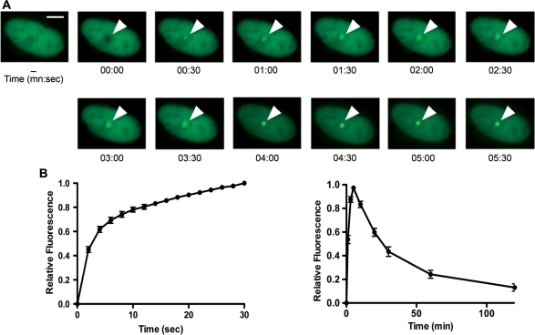
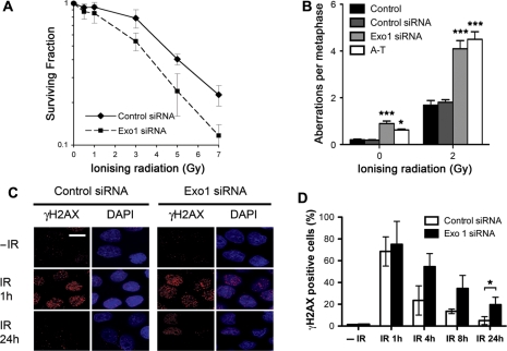
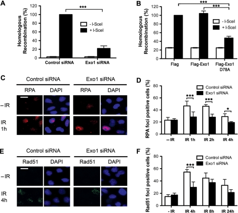
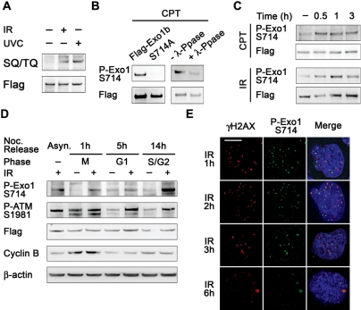
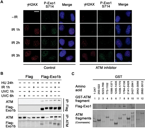
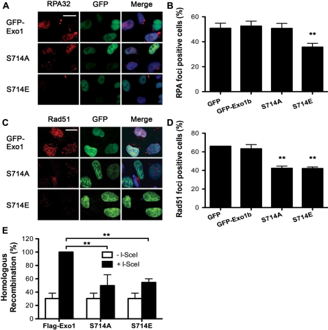
DNA double-strand break (DSB) repair via the homologous recombination pathway is a multi-stage process, which results in repair of the DSB without loss of genetic information or fidelity. One essential step in this process is the generation of extended single-stranded DNA (ssDNA) regions at the break site. This ssDNA serves to induce cell cycle checkpoints and is required for Rad51 mediated strand invasion of the sister chromatid. Here, we show that human Exonuclease 1 (Exo1) is required for the normal repair of DSBs by HR. Cells depleted of Exo1 show chromosomal instability and hypersensitivity to ionising radiation (IR) exposure. We find that Exo1 accumulates rapidly at DSBs and is required for the recruitment of RPA and Rad51 to sites of DSBs, suggesting a role for Exo1 in ssDNA generation. Interestingly, the phosphorylation of Exo1 by ATM appears to regulate the activity of Exo1 following resection, allowing optimal Rad51 loading and the completion of HR repair. These data establish a role for Exo1 in resection of DSBs in human cells, highlighting the critical requirement of Exo1 for DSB repair via HR and thus the maintenance of genomic stability.


Figures

Figure 1.
Figure 1.
Recruitment of Exo1 to DNA DSBs. (A) U2OS cells expressing GFP-Exo1b fusion protein were micro-irradiated and images taken at the indicated times after damage. The scale bar represents 5 µm. (B) Graphical representation of Exo1 recruitment to sites of DNA damage from 0 to 30 s after damage induction. (C) Graphical representation of Exo1 recruitment and dissociation from sites of DNA damage from 0 to 120 min after damage induction. The data shown represents the mean and standard deviation (SD) of three independent experiments.
Figure 2.
Figure 2.
DNA repair defects in Exo1-depleted cells. (A) Colony survival in HeLa cells transfected with control and Exo1 siRNA and treated with the indicated dose of IR. (B) Frequencies of spontaneous and IR (2 Gy) induced chromosome and chromatid breaks and gaps in control and Exo1-deficient cells are indicated. Chromosomal fragments are consequences of breaks either at the chromosome level or at the chromatid level. A-T cells are used as positive control. Fifty metaphases for each sample were analyzed. (C) and (D) HeLa cells were transfected with control or Exo1 siRNA and treated with 2 Gy IR before fixation and staining with γH2AX antibodies at the indicated times. Representative images (C) and graphical representation of data (D) are shown. The scale bar represents 10 µm. The results shown are the average of three independent experiments and error bars indicate the SD. One star and three stars designate statistical significance in a two-way ANOVA. *P < 0.05 and ***P < 0.001.
Figure 3.
Figure 3.
Recruitment and repair defects in Exo1-deficient cells. (A) HR repair is reduced in cells transfected with Exo1 siRNA. Twenty-four hours after siRNA transfection MCF7-DRGFP cells were transfected with I-SceI plasmid (pCBSCE). Fourty-eight hours after pCBSCE transfection FACS analysis was carried out to detect GFP positive cells. (B) HR repair is reduced in cells expressing nuclease-deficient Exo1. Twenty-four hours after transfection with empty vector, wild-type Exo1 or D78A, MCF7-DRGFP cells were transfected with I-SceI plasmid (as above). Forty-eight hours after pCBSCE transfection FACS analysis was carried out to detect GFP positive cells. The results shown are the average of three independent experiments and error bars indicate the SD (A) and (B). (C)–(F) HeLa cells were transfected with control or Exo1 siRNA and treated with 5 Gy IR before fixation and staining with RPA34 (C) and (D) or Rad51 (E) and (F) antibodies. The scale bar represents 10 µm. The mean and SD from two-four independent experiments is depicted (D) and (F). One star and three stars designate statistical significance in a two-way ANOVA. *P < 0.05 and ***P < 0.001.
Figure 4.
Figure 4.
Exo1 is phosphorylated on S714 after DSB induction. (A) HeLa cells were transfected with Flag-Exo1 and treated with 6 Gy IR or 40 J/m2 UVC and extracts taken after 1 h. Extracts were immunoprecipitated with Flag M2 beads and immunoblotted with an antibody that specifically recognises phosphorylated SQ/TQ residues and Flag antibodies. (B) HeLa cells were transfected with Flag-Exo1 or Flag-S714A and treated with 1 µM CPT; extracts were taken after 1 h. Extracts were immunoprecipitated [as in (A)] and immunoblotted with the indicated antibodies. Second panel, cell extracts were incubated in the presence of λ-phosphatase where indicated. Immunoprecipitates were immunoblotted with the indicated antibodies. (C) HeLa cells were transfected with Flag-Exo1 and treated with 1 µM CPT (top panels) or 6 Gy IR (bottom panels). Extracts were taken after the indicated time and immunoprecipitated as in (B). Immunoprecipitates were immunoblotted with the indicated antibodies. (D) HeLa cells were transfected with Flag-Exo1 and synchronised using nocodazole. Cells were treated with 6 Gy IR at the indicated stage of the cell cycle and extracts were taken after 1 h. Extracts were immunoblotted with the indicated antibodies. (E) HeLa cells were treated with 1 Gy IR and fixed after 1 h. Immunofluorescence was carried out using the indicated antibodies. The scale bar represents 5 µm.
Figure 5.
Figure 5.
Exo1 interacts with ATM and phosphorylation of Exo1 on S714 is dependent upon ATM. (A) HeLa cells were pretreated or mock-treated with the ATM inhibitor, KU55933 (10 µM) 1 h prior to treatment with 6 Gy IR and fixed at the indicated time points. Cells were immunostained with the indicated antibodies. The scale bar represents 10 µm. (B) Top panels, HeLa cells were transfected with Flag-Exo1 and treated with 40 J/M2 UVC, 6 Gy IR or 2 mM HU and extracts taken after the indicated time. Immunoprecipitations were carried out using Flag M2 Beads. Immunoprecipitates were immunoblotted with the indicated antibodies. Bottom panels, HeLa cells were treated as above and immunoprecipitated using ATM antibodies. Immunoprecipitates were immunoblotted with the indicated antibodies. (C) Exo1 interacts with GST-ATM fragment 8 (amino acids 1764–2138). Recombinant GST-ATM fragments representing the full length of ATM were used in pull-down assays. Total cell extracts from HeLa cells expressing Flag-Exo1 were mixed with glutathione agarose beads containing GST-ATM fusion proteins. Bound proteins were analysed by immunoblotting with Flag antibodies (top panel) and levels of GST-ATM fragments were detected by coomassie staining (bottom panel).
Figure 6.
Figure 6.
Exo1 phosphorylation is required for recruitment of Rad51 and DSB repair by HR. (A) and (B) HeLa cells were transfected with the indicated constructs. Cells were treated with 5 Gy IR and fixed 2-h after treatment. Immunofluorescence was carried out using RPA34 antibodies. Cells expressing GFP were scored as positive or negative for RPA34 foci. Representative images are shown. Two stars designate significant statistical difference to the control in a t-test, **P = 0.0032. (C) and (D) HeLa cells were transfected with the indicated constructs. Cells were treated with 5 Gy IR and fixed 4-h after treatment. Immunofluorescence was carried out using Rad51 antibodies. Cells expressing GFP were scored as positive or negative for Rad51 foci. Representative images are shown. Two stars designate significant statistical difference to the control in a t-test, **P < 0.005. The scale bar represents 10 µm. (E) HR repair is reduced in cells expressing S714A and S714E. Twenty-four hours after transfection with wild-type Exo1, S714A or S714E, MCF7-DRGFP cells were transfected with I-SceI plasmid (pCBSCE). Forty-eight hours after pCBSCE transfection FACS analysis was carried out to detect GFP positive cells. Two stars designate statistical significance in a two-way ANOVA. **P < 0.001. (B), (D) and (E) The results shown are the average of three independent experiments and error bars indicate the SD.

Similar articles

Cited by

References

    1. Mimitou EP, Symington LS. Nucleases and helicases take center stage in homologous recombination. Trends Biochem. Sci. 2009;34:264–272. - PubMed
    1. Shiotani B, Zou L. Single-stranded DNA orchestrates an ATM-to-ATR switch at DNA breaks. Mol. Cell. 2009;33:547–558. - PMC - PubMed
    1. Sartori AA, Lukas C, Coates J, Mistrik M, Fu S, Bartek J, Baer R, Lukas J, Jackson SP. Human CtIP promotes DNA end resection. Nature. 2007;450:509–514. - PMC - PubMed
    1. Bernstein KA, Rothstein R. At loose ends: resecting a double-strand break. Cell. 2009;137:807–810. - PMC - PubMed
    1. Szankasi P, Smith GR. A DNA exonuclease induced during meiosis of Schizosaccharomyces pombe. J. Biol. Chem. 1992;267:3014–3023. - PubMed

Publication types

MeSH terms