. 2010 Jan;21(1):124-35.
doi: 10.1681/ASN.2008101059. Epub 2009 Dec 17.

Dickkopf-1 promotes hyperglycemia-induced accumulation of mesangial matrix and renal dysfunction

Affiliations

Dickkopf-1 promotes hyperglycemia-induced accumulation of mesangial matrix and renal dysfunction

Chun-Liang Lin et al. J Am Soc Nephrol. 2010 Jan.

Abstract

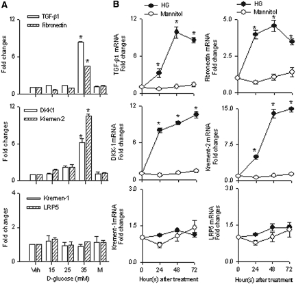
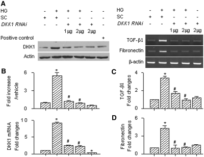
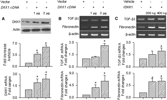
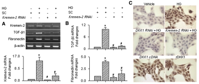
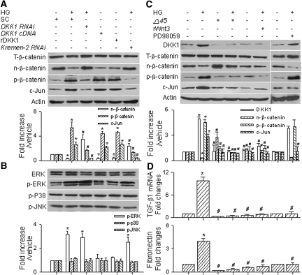
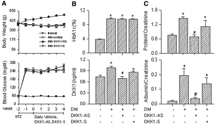
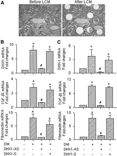
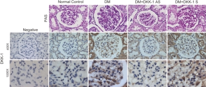
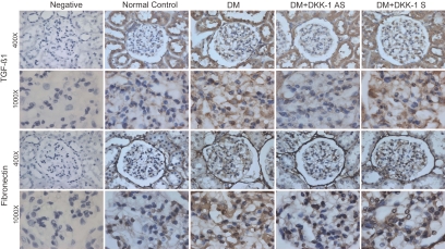
Wnt/beta-catenin signaling mediates renal fibrosis in several model systems including diabetic nephropathy. Dickkopf-1 (DKK-1) is an endogenous inhibitor of Wnt/beta-catenin signaling, but whether DKK-1 modulates diabetic nephropathy is unknown. Here, we studied whether DKK-1 participates in high glucose (HG)-induced expression of profibrotic factors and renal damage. In vitro, HG increased expression of DKK1, receptor Kremen-2, TGF-beta1, and fibronectin in mesangial cells. Loss and gain of DKK1 function modulated HG-mediated c-Jun, TGF-beta1, and fibronectin expression. DKK1 mediated HG-induced phosphorylation of Ser45-beta-catenin and reduction of nuclear beta-catenin levels, but not phosphorylation of ERK kinase. Wnt3a protein and the beta-catenin (Delta45) mutation increased nuclear beta-catenin but abrogated HG-induced DKK1 and fibronectin expression. Exogenous DKK1 antisense oligonucleotide attenuated the increase in both serum DKK1 and urinary protein excretion in streptozotocin-induced diabetic rats. Knocking down DKK1 inhibited mesangial expression of TGF-beta1 and fibronectin and reduced both the glomerular volume and deposition of mesangial matrix in diabetic kidneys. Taken together, DKK1 mediates HG-induced destabilization of beta-catenin and matrix accumulation in mesangial cells. Knocking down DKK1 prevents diabetes-induced renal dysfunction and microstructure deterioration, suggesting that inhibition of DKK1offers therapeutic potential for diabetic nephropathy.


Figures

Figure 1.
Figure 1.
Concentration and time effects of d-glucose on expression of DKK1 and profibrotic factor in renal mesangial cells. (A) Increased d-glucose concentration augmented expression of TGF-β1 and fibronectin in association with increased expression of DKK1 and Kremen-2 in cell cultures. d-Glucose at 35 mM had the highest effect on mRNA expression in cell cultures. (B) d-Glucose at 35 mM increased TGF-β1, fibronectin, DKK1, and Kremen-2 expression by 24 hours. Increased d-glucose did not significantly affect Kremen-1 or LRP5 mRNA expression throughout the study period. Cells (1 × 10⁶ cell/well, in a six-well plate) were cultured in medium containing 15 to 35 mM d-glucose or the osmolarity control 35 mM mannitol for 24, 48, and 72 hours. The graphed results represent the relative abundance of mRNAs determined by quantitative RT-PCR and normalized to the housekeeping gene β-actin. Experimental results are presented as means ± SEs calculated from at least three experiments. *Significant difference (P < 0.05) from the vehicle groups. Veh, vehicle; M, 35 mM mannitol.
Figure 2.
Figure 2.
Effect of DKK1 RNAi on expression of fibrotic factor in mesangial cells. (A through D) DKK1 RNAi significantly attenuated expression of DKK1 protein (A), DKK1 mRNA (B), TGF-β1 (C), and fibronectin mRNA (D) induced by HG in cell cultures. Mesangial cells transfected with DKK1 siRNA and scrambled control were cultured in HG for 72 hours. The graphed results represent relative abundances of DKK1, TGF-β1, and fibronectin mRNA determined by real-time PCR and normalized to the housekeeping gene β-actin. Immunoblotting for actin showed equal loading and transfer for all lanes. Experimental results are presented as means ± SEs calculated from at least three experiments. *, #Significant differences (P < 0.05) from the vehicle- and HG-treated groups, respectively. SC, scramble control.
Figure 3.
Figure 3.
Effects of DKK1 cDNA and recombinant DKK1 protein on expression of profibrotic factor in mesangial cells. (A and B) DKK1 cDNA increased DKK1 protein and mRNA expression (A) and promoted TGF-β1 and fibronectin mRNA expression (B) in cell cultures. (C) Treatment with recombinant DKK1 protein induced TGF-β1 and fibronectin expression in mesangial cells. Mesangial cells transfected with DKK1 cDNA and empty vector were cultured in basal medium for 72 hours. Cell cultures were treated with 200 and 400 ng/ml recombinant DKK1 protein for 72 hours. Experimental results are presented as means ± SEs calculated from at least three experiments. *Significant differences (P < 0.05) from the vehicle groups. rDKK1, recombinant DKK1 protein.
Figure 4.
Figure 4.
Effect of Kremen-2 RNAi on expression of fibrotic factor in mesangial cells. (A and B) Kemen-2 RNAi attenuated expression of Kremen-2 (A) and TGF-β1 and fibronectin (B) induced by HG in mesangial cell cultures. (C) Representative immunocytochemical photographs of mesangial cells. In the HG and DKK1 cDNA groups, mesangial cells displayed intense fibronectin expression. Cell cultures treated with DKK1 RNAi and Kremen-2 RNAi showed weak fibronectin expression. Expression of fibronectin in cell cultures was determined immunocytochemically with anti-fibronectin antibody and horseradish peroxidase-3′-3′-diaminobenzidene. Specimens were observed under ×400 magnification. Experimental results are presented as means ± SEs calculated from at least three experiments. *, #Significant differences (P < 0.05) from the vehicle- and HG-treated groups, respectively. SC, scramble control; rDKK1, recombinant DKK1 protein.
Figure 5.
Figure 5.
Effects of treatment with HG, DKK1, and Wnt on expression of β-catenin and mitogen-activated protein kinase in mesangial cells. (A) HG, DKK1 cDNA, and recombinant DKK1 protein (400 ng/ml) increased expression of phosphorylated Ser45–β-catenin and nuclear c-Jun but inhibited nuclear β-catenin expression. DKK1 and Kremen-2 siRNA abrogated expression of phosphorylated Ser45–β-catenin and c-Jun but increased nuclear β-catenin induced by HG. (B) Controlling DKK1 and Kremen-2 did not markedly alter expression of phosphorylated ERK, phosphorylated p38, or phosphorylated JNK in cell cultures. (C) Recombinant Wnt3a protein and the β-catenin (Δ45) mutation alleviated expression of phosphorylated Ser45–β-catenin and c-Jun but attenuated expression of nuclear β-catenin induced by HG. (D) Recombinant Wnt3a protein and the β-catenin (Δ45) mutation abrogated expression of TGF-β1 and fibronectin mRNA in cell cultures. Pretreatment with PD98059 did not alter DKK1 and β-catenin expression but attenuated expression of c-Jun, TGF-β1, and fibronectin induced by HG in cell cultures. Immunoblotting for actin and ERK showed equal loading and transfer for all lanes. *, #Significant differences (P < 0.05) from the vehicle- and HG-treated groups, respectively. SC, scramble control; rDKK1, recombinant DKK1 protein; T-β-catenin, total β-catenin; n-β-catenin, nuclear β-catenin; p-β-catenin, phosphorylated Ser45–β-catenin; p-ERK, phosphorylated ERK; p-p38, phosphorylated p38; p-JNK, phosphorylated JNK.
Figure 6.
Figure 6.
Effect of DKK1 antisense oligonucleotide on renal function in diabetic rats. (A) Diabetes attenuated body weight but increased blood glucose levels in rats. Diabetes increased levels of HbA1c and serum DKK1 (B) and urinary protein and albumin (C) in experimental animals. Treatment with exogenous DKK1-AS attenuated levels of serum DKK1 and urinary protein and albumin in diabetic rats. Rats were given STZ to induce diabetes. Two weeks after injection, diabetic rats were given 20 μg/kg per d DKK1-AS (n = 6), DKK1-S (n = 6), and vehicle (n = 6) for 4 consecutive weeks. Experimental results are presented as means ± SEs calculated from six rats in each group. *, #Significant differences (P < 0.05) from the normal control and diabetic groups, respectively. DM, diabetes.
Figure 7.
Figure 7.
Effect of treatment with DKK1 antisense oligonucleotide on expression of DKK1 and fibrotic factor in whole renal tissue and glomerular mesangium from diabetic kidneys. (A) Representative histologic photographs of renal tissue before and after dissection of the glomerular mesangium. DKK1-AS attenuated expression of DKK1, TGF-β1, and fibronectin induced by diabetes in whole renal tissue (B) and the glomerular mesangium (C). The glomerular mesangium in rat kidneys was harvested by laser-captured microdissection. The graphed results represent the relative abundances of DKK1, TGF-β1, and fibronectin mRNA determined by real-time PCR and normalized to the housekeeping gene β-actin. Experimental results are presented as means ± SEs calculated from six rats. *, #Significant differences (P < 0.05) from the vehicle- and HG-treated groups, respectively. DM, diabetes; LCM, laser-captured microdissection.
Figure 8.
Figure 8.
Representative histologic photographs of glomeruli in diabetic kidneys. In the diabetes and DKK1-S groups, glomeruli displayed intense PAS staining and DKK1 expression. In the DKK1-AS and normal groups, glomeruli displayed weak PAS staining. Mesangial cells and podocytes in glomeruli and tubular cells expressed DKK1 weakly. Cells positively immunostained for DKK1 showed brown color. Specimens were observed under ×400 and ×1000 magnification. DM, diabetes.
Figure 9.
Figure 9.
Representative photographs of β-catenin and c-Jun immunostaining in glomeruli from diabetic kidneys. In the DKK1-S and diabetes groups, mesangial cells, podocytes, and tubular cells expressed β-catenin weakly but showed intense immunostaining for c-Jun. In the DKK1-AS and normal groups, the cells displayed weak c-Jun immunoreactivity but strong β-catenin expression. Cells positively immunostained for c-Jun and β-catenin showed brown color. Specimens were observed under ×400 and ×1000 magnification. DM, diabetes.
Figure 10.
Figure 10.
Representative photographs of immunostaining for TGF-β1 and fibronectin in glomeruli from diabetic kidneys. In the DKK1-S and diabetes groups, mesangial cells, podocytes, and tubular cells displayed strong TGF-β1, fibronectin, and β-catenin expression. In the DKK1-AS and normal groups, the cells displayed weak TGF-β1 and fibronectin immunoreactivity. Cells positively immunostained for TGF-β1 and fibronectin showed brown color. Specimens were observed under ×400 and ×1000 magnification. DM, diabetes.

References

    1. Simonson MS: Phenotypic transitions and fibrosis in diabetic nephropathy. Kidney Int 71: 846–854, 2007 - PubMed
    1. Mizuno S, Nakamura T: Suppressions of chronic glomerular injuries and TGF-beta 1 production by HGF in attenuation of murine diabetic nephropathy. Am J Physiol Renal Physiol 286: F134–F143, 2004 - PubMed
    1. Furlong F, Crean J, Thornton L, O'Leary R, Murphy M, Martin F: Dysregulated intracellular signaling impairs CTGF-stimulated responses in human mesangial cells exposed to high extracellular glucose. Am J Physiol Renal Physiol 292: F1691–F1700, 2007 - PubMed
    1. Giannico G, Cortes P, Baccora MH, Hassett C, Taube DW, Yee J: Glibenclamide prevents increased extracellular matrix formation induced by high glucose concentration in mesangial cells. Am J Physiol Renal Physiol 292: F57–F65, 2007 - PubMed
    1. Lee EA, Seo JY, Jiang Z, Yu MR, Kwon MK, Ha H, Lee HB: Reactive oxygen species mediate high glucose-induced plasminogen activator inhibitor-1 up-regulation in mesangial cells and in diabetic kidney. Kidney Int 67: 1762–1771, 2005 - PubMed

Publication types

MeSH terms