

New insights into epithelial-mesenchymal transition in kidney fibrosis
- PMID: 20019167
- PMCID: PMC4554339
- DOI: 10.1681/ASN.2008121226
New insights into epithelial-mesenchymal transition in kidney fibrosis
Abstract
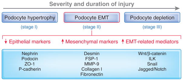
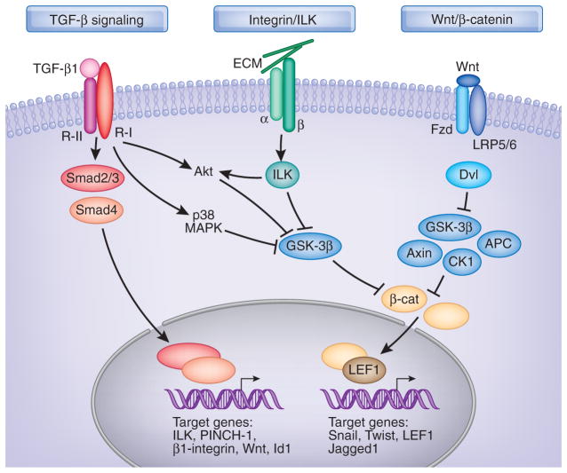
Epithelial-mesenchymal transition (EMT), a process by which differentiated epithelial cells undergo a phenotypic conversion that gives rise to the matrix-producing fibroblasts and myofibroblasts, is increasingly recognized as an integral part of tissue fibrogenesis after injury. However, the degree to which this process contributes to kidney fibrosis remains a matter of intense debate and is likely to be context-dependent. EMT is often preceded by and closely associated with chronic interstitial inflammation and could be an adaptive response of epithelial cells to a hostile or changing microenvironment. In addition to tubular epithelial cells, recent studies indicate that endothelial cells and glomerular podocytes may also undergo transition after injury. Phenotypic alteration of podocytes sets them in motion to functional impairment, resulting in proteinuria and glomerulosclerosis. Several intracellular signal transduction pathways such as TGFbeta/Smad, integrin-linked kinase (ILK) and Wnt/beta-catenin signaling are essential in controlling the process of EMT and presently are potential targets of antifibrotic therapy. This review highlights the current understanding of EMT and its underlying mechanisms to stimulate further discussion on its role, not only in the pathogenesis of renal interstitial fibrosis but also in the onset of podocyte dysfunction, proteinuria, and glomerulosclerosis.
Figures
Similar articles
-
Epithelial to mesenchymal transition in renal fibrogenesis: pathologic significance, molecular mechanism, and therapeutic intervention.J Am Soc Nephrol. 2004 Jan;15(1):1-12. doi: 10.1097/01.asn.0000106015.29070.e7. J Am Soc Nephrol. 2004. PMID: 14694152 Review.
-
Role for integrin-linked kinase in mediating tubular epithelial to mesenchymal transition and renal interstitial fibrogenesis.J Clin Invest. 2003 Aug;112(4):503-16. doi: 10.1172/JCI17913. J Clin Invest. 2003. PMID: 12925691 Free PMC article.
-
New insights into the regulation of epithelial-mesenchymal transition and tissue fibrosis.Int Rev Cell Mol Biol. 2012;294:171-221. doi: 10.1016/B978-0-12-394305-7.00004-5. Int Rev Cell Mol Biol. 2012. PMID: 22364874 Review.
-
Renal interstitial fibrosis: a critical evaluation of the origin of myofibroblasts.Contrib Nephrol. 2011;169:73-93. doi: 10.1159/000313946. Epub 2011 Jan 20. Contrib Nephrol. 2011. PMID: 21252512
-
Epithelial-mesenchymal transition in renal fibrosis - evidence for and against.Int J Exp Pathol. 2011 Jun;92(3):143-50. doi: 10.1111/j.1365-2613.2011.00775.x. Epub 2011 May 6. Int J Exp Pathol. 2011. PMID: 21554437 Free PMC article. Review.
Cited by
-
Histone methylation modification and diabetic kidney disease: Potential molecular mechanisms and therapeutic approaches (Review).Int J Mol Med. 2024 Nov;54(5):104. doi: 10.3892/ijmm.2024.5428. Epub 2024 Sep 20. Int J Mol Med. 2024. PMID: 39301658 Free PMC article. Review.
-
Selective activation of p120ctn-Kaiso signaling to unlock contact inhibition of ARPE-19 cells without epithelial-mesenchymal transition.PLoS One. 2012;7(5):e36864. doi: 10.1371/journal.pone.0036864. Epub 2012 May 9. PLoS One. 2012. PMID: 22590627 Free PMC article.
-
Severe intraglomerular detachment of podocytes in a Gitelman syndrome patient.Clin Exp Nephrol. 2012 Jun;16(3):495-500. doi: 10.1007/s10157-012-0624-4. Epub 2012 Apr 7. Clin Exp Nephrol. 2012. PMID: 22484642
-
Cellular and Molecular Mechanisms of Chronic Kidney Disease with Diabetes Mellitus and Cardiovascular Diseases as Its Comorbidities.Front Immunol. 2015 Jul 8;6:340. doi: 10.3389/fimmu.2015.00340. eCollection 2015. Front Immunol. 2015. PMID: 26217336 Free PMC article. Review.
-
Matrix metalloproteinases in kidney homeostasis and diseases.Am J Physiol Renal Physiol. 2012 Jun 1;302(11):F1351-61. doi: 10.1152/ajprenal.00037.2012. Epub 2012 Apr 4. Am J Physiol Renal Physiol. 2012. PMID: 22492945 Free PMC article. Review.
References
-
- Eddy AA. Molecular basis of renal fibrosis. Pediatr Nephrol. 2000;15:290–301. - PubMed
-
- Thiery JP. Epithelial-mesenchymal transitions in development and pathologies. Curr Opin Cell Biol. 2003;15:740–746. - PubMed
-
- Liu Y. Epithelial to mesenchymal transition in renal fibrogenesis: Pathologic significance, molecular mechanism, and therapeutic intervention. J Am Soc Nephrol. 2004;15:1–12. - PubMed
Publication types
MeSH terms
Grants and funding
LinkOut - more resources
Full Text Sources
