

Reduced expression of integrin alphavbeta8 is associated with brain arteriovenous malformation pathogenesis
- PMID: 20019187
- PMCID: PMC2808104
- DOI: 10.2353/ajpath.2010.090453
Reduced expression of integrin alphavbeta8 is associated with brain arteriovenous malformation pathogenesis
Abstract
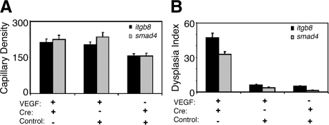
Brain arteriovenous malformations (BAVMs) are a rare but potentially devastating hemorrhagic disease. Transforming growth factor-beta signaling is required for proper vessel development, and defective transforming growth factor-beta superfamily signaling has been implicated in BAVM pathogenesis. We hypothesized that expression of the transforming growth factor-beta activating integrin, alphavbeta8, is reduced in BAVMs and that decreased beta8 expression leads to defective neoangiogenesis. We determined that beta8 protein expression in perivascular astrocytes was reduced in human BAVM lesional tissue compared with controls and that the angiogenic response to focal vascular endothelial growth factor stimulation in adult mouse brains with local Cre-mediated deletion of itgb8 and smad4 led to vascular dysplasia in newly formed blood vessels. In addition, common genetic variants in ITGB8 were associated with BAVM susceptibility, and ITGB8 genotypes associated with increased risk of BAVMs correlated with decreased beta8 immunostaining in BAVM tissue. These three lines of evidence from human studies and a mouse model suggest that reduced expression of integrin beta8 may be involved in the pathogenesis of sporadic BAVMs.
Figures


Similar articles
-
Integrin β8 Deletion Enhances Vascular Dysplasia and Hemorrhage in the Brain of Adult Alk1 Heterozygous Mice.Transl Stroke Res. 2016 Dec;7(6):488-496. doi: 10.1007/s12975-016-0478-2. Epub 2016 Jun 29. Transl Stroke Res. 2016. PMID: 27352867 Free PMC article.
-
Mesenchymal Behavior of the Endothelium Promoted by SMAD6 Downregulation Is Associated With Brain Arteriovenous Malformation Microhemorrhage.Stroke. 2020 Jul;51(7):2197-2207. doi: 10.1161/STROKEAHA.120.030046. Epub 2020 Jun 3. Stroke. 2020. PMID: 32486965
-
Reductions in brain pericytes are associated with arteriovenous malformation vascular instability.J Neurosurg. 2018 Dec 1;129(6):1464-1474. doi: 10.3171/2017.6.JNS17860. Epub 2018 Jan 5. J Neurosurg. 2018. PMID: 29303444 Free PMC article.
-
Brain arteriovenous malformation modeling, pathogenesis, and novel therapeutic targets.Transl Stroke Res. 2014 Jun;5(3):316-29. doi: 10.1007/s12975-014-0343-0. Epub 2014 Apr 12. Transl Stroke Res. 2014. PMID: 24723256 Free PMC article. Review.
-
Pathogenesis and radiobiology of brain arteriovenous malformations: implications for risk stratification in natural history and posttreatment course.Neurosurg Focus. 2009 May;26(5):E9. doi: 10.3171/2009.2.FOCUS0926. Neurosurg Focus. 2009. PMID: 19409010 Review.
Cited by
-
An early venous abnormality: a potential cause of arteriovenous malformation recurrence.BMJ Case Rep. 2012 Sep 11;2012:bcr0120125646. doi: 10.1136/bcr.01.2012.5646. BMJ Case Rep. 2012. PMID: 22967676 Free PMC article.
-
Managing MMP-2, MMP-9, VEGFR-2, TGFβ-1, and TIMP-1 in NNK-induced lung carcinoma by nonchemical interventions in female rats.Toxicol Rep. 2022 May 30;9:1261-1267. doi: 10.1016/j.toxrep.2022.05.018. eCollection 2022. Toxicol Rep. 2022. PMID: 36518397 Free PMC article.
-
Shaping the brain vasculature in development and disease in the single-cell era.Nat Rev Neurosci. 2023 May;24(5):271-298. doi: 10.1038/s41583-023-00684-y. Epub 2023 Mar 20. Nat Rev Neurosci. 2023. PMID: 36941369 Free PMC article. Review.
-
Genome-wide association study of sporadic brain arteriovenous malformations.J Neurol Neurosurg Psychiatry. 2016 Sep;87(9):916-23. doi: 10.1136/jnnp-2015-312272. Epub 2016 Jan 27. J Neurol Neurosurg Psychiatry. 2016. PMID: 26818729 Free PMC article.
-
Single Cell RNA Sequencing and Spatial Profiling Identify Mechanisms of Neonatal Brain Hemorrhage Development and Resolution.bioRxiv [Preprint]. 2025 Jul 31:2025.07.30.667675. doi: 10.1101/2025.07.30.667675. bioRxiv. 2025. PMID: 40766655 Free PMC article. Preprint.
References
-
- Young WL, Kwok PY, Pawlikowska L, Lawton MT, Kim H, Hysi PG, Marchuk DA. Arteriovenous malformation. J Neurosurg. 2007;106:731–733. - PubMed
-
- Chen Y, Fan Y, Poon KY, Achrol AS, Lawton MT, Zhu Y, McCulloch CE, Hashimoto T, Lee C, Barbaro NM, Bollen AW, Yang GY, Young WL. MMP-9 expression is associated with leukocytic but not endothelial markers in brain arteriovenous malformations. Front Biosci. 2006;11:3121–3128. - PubMed
-
- Kim H, Pawlikowska L, Young WL: Molecular and genetic aspects of brain vascular malformations. Edited by Mohr JP, Wolf PA, Grotta JC, Moskowitz MA, Mayberg M, von Kummer R. Philadelphia, Churchill Livingstone Elsevier (in press)
-
- Young WL, Yang GY. Are there genetic influences on sporadic brain arteriovenous malformations? Stroke. 2004;35:2740–2745. - PubMed
Publication types
MeSH terms
Substances
Grants and funding
LinkOut - more resources
Full Text Sources
Molecular Biology Databases
Miscellaneous
