

Human nephrosclerosis triggers a hypoxia-related glomerulopathy
- PMID: 20019191
- PMCID: PMC2808068
- DOI: 10.2353/ajpath.2010.090268
Human nephrosclerosis triggers a hypoxia-related glomerulopathy
Abstract
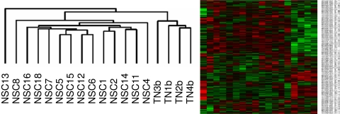
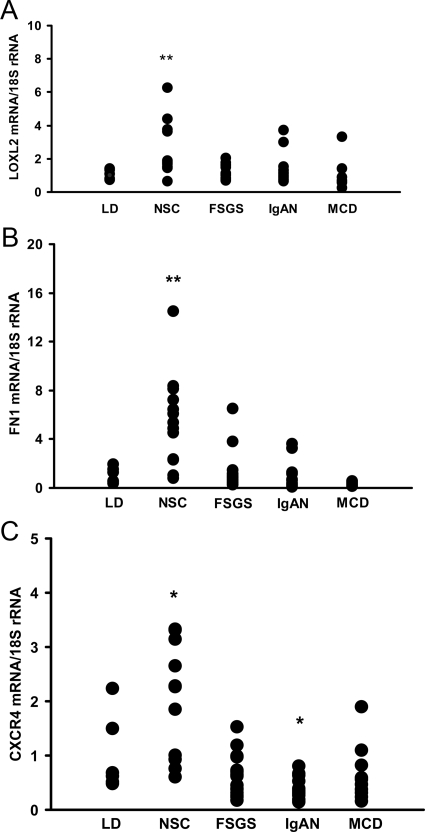
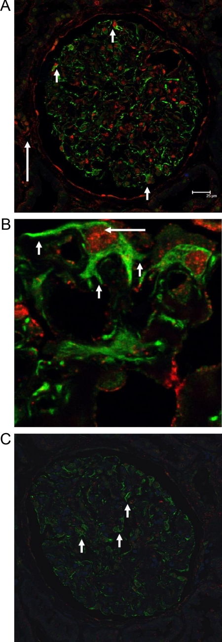
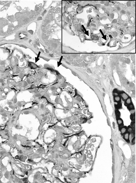
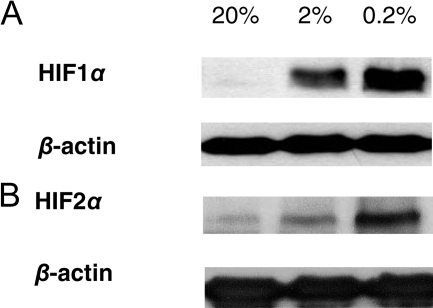
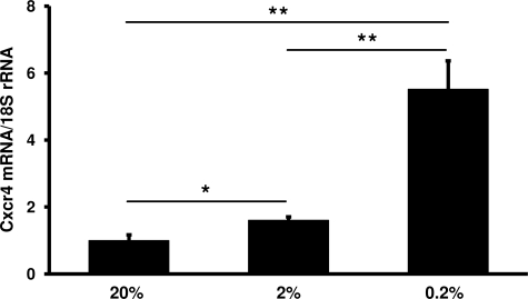
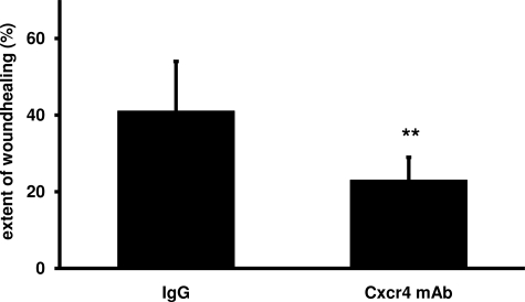
In the kidney, hypoxia contributes to tubulointerstitial fibrosis, but little is known about its implications for glomerular damage and glomerulosclerosis. Chronic hypoxia was hypothesized to be involved in nephrosclerosis (NSC) or "hypertensive nephropathy." In the present study genome-wide expression data from microdissected glomeruli were studied to examine the role of hypoxia in glomerulosclerosis of human NSC. Functional annotation analysis revealed prominent regulation of hypoxia-associated biological processes in NSC, including angiogenesis, fibrosis, and inflammation. Glomerular expression levels of a majority of genes regulated by the hypoxia-inducible factors (HIFs) were significantly altered in NSC. Among these HIF targets, chemokine C-X-C motif receptor 4 (CXCR4) was prominently induced. Glomerular CXCR4 mRNA induction was confirmed by quantitative RT-PCR in an independent cohort with NSC but not in those with other glomerulopathies. By immunohistological analysis, CXCR4 showed enhanced positivity in podocytes in NSC biopsy specimens. This CXCR4 positivity was associated with nuclear localization of HIF1alpha only in podocytes of NSC, indicating transcriptional activity of HIF. As the CXCR4 ligand CXCL12/SDF-1 is constitutively expressed in podocytes, autocrine signaling may contribute to NSC. In addition, a blocking CXCR4 antibody caused significant inhibition of wound closure by podocytes in an in vitro scratch assay. These data support a role for CXCR4/CXCL12 in human NSC and indicate that hypoxia not only is involved in tubulointerstitial fibrosis but also contributes to glomerular damage in NSC.
Figures
Similar articles
-
Stabilization of hypoxia-inducible factor ameliorates glomerular injury sensitization after tubulointerstitial injury.Kidney Int. 2021 Mar;99(3):620-631. doi: 10.1016/j.kint.2020.09.031. Epub 2020 Nov 1. Kidney Int. 2021. PMID: 33137336 Free PMC article.
-
Podocyte loss in human hypertensive nephrosclerosis.Am J Hypertens. 2009 Mar;22(3):300-6. doi: 10.1038/ajh.2008.360. Epub 2009 Jan 8. Am J Hypertens. 2009. PMID: 19131934
-
The CXCR4 antagonist AMD3100 suppresses hypoxia-mediated growth hormone production in GH3 rat pituitary adenoma cells.J Neurooncol. 2010 Oct;100(1):51-64. doi: 10.1007/s11060-010-0152-6. Epub 2010 Mar 23. J Neurooncol. 2010. PMID: 20309720
-
Detrimental effects of hypoxia on glomerular podocytes.J Physiol Biochem. 2021 May;77(2):193-203. doi: 10.1007/s13105-021-00788-y. Epub 2021 Apr 9. J Physiol Biochem. 2021. PMID: 33835424 Review.
-
[Mechanisms in remodeling of the kidney].Nihon Rinsho. 2008 Sep;66(9):1696-700. Nihon Rinsho. 2008. PMID: 18788396 Review. Japanese.
Cited by
-
Lysyl Oxidase-like-2 Cross-links Collagen IV of Glomerular Basement Membrane.J Biol Chem. 2016 Dec 9;291(50):25999-26012. doi: 10.1074/jbc.M116.738856. Epub 2016 Oct 21. J Biol Chem. 2016. PMID: 27770022 Free PMC article.
-
BAMBI is expressed in endothelial cells and is regulated by lysosomal/autolysosomal degradation.PLoS One. 2010 Sep 24;5(9):e12995. doi: 10.1371/journal.pone.0012995. PLoS One. 2010. PMID: 20886049 Free PMC article.
-
Aristolochic acid I and ochratoxin A differentially regulate VEGF expression in porcine kidney epithelial cells--the involvement of SP-1 and HIFs transcription factors.Toxicol Lett. 2011 Jul 28;204(2-3):118-26. doi: 10.1016/j.toxlet.2011.04.022. Epub 2011 Apr 29. Toxicol Lett. 2011. PMID: 21554934 Free PMC article.
-
A Functional Landscape of CKD Entities From Public Transcriptomic Data.Kidney Int Rep. 2019 Nov 13;5(2):211-224. doi: 10.1016/j.ekir.2019.11.005. eCollection 2020 Feb. Kidney Int Rep. 2019. PMID: 32043035 Free PMC article.
-
Periostin is induced in glomerular injury and expressed de novo in interstitial renal fibrosis.Am J Pathol. 2011 Oct;179(4):1756-67. doi: 10.1016/j.ajpath.2011.06.002. Epub 2011 Aug 18. Am J Pathol. 2011. PMID: 21854746 Free PMC article.
References
Publication types
MeSH terms
Substances
Grants and funding
LinkOut - more resources
Full Text Sources
Medical
Molecular Biology Databases
Research Materials
