. 2010 Jan 29;327(5965):555-9.
doi: 10.1126/science.1182340. Epub 2009 Dec 17.

Spontaneous and x-ray-triggered crystallization at long range in self-assembling filament networks

Affiliations

Spontaneous and x-ray-triggered crystallization at long range in self-assembling filament networks

Honggang Cui et al. Science. .

Abstract

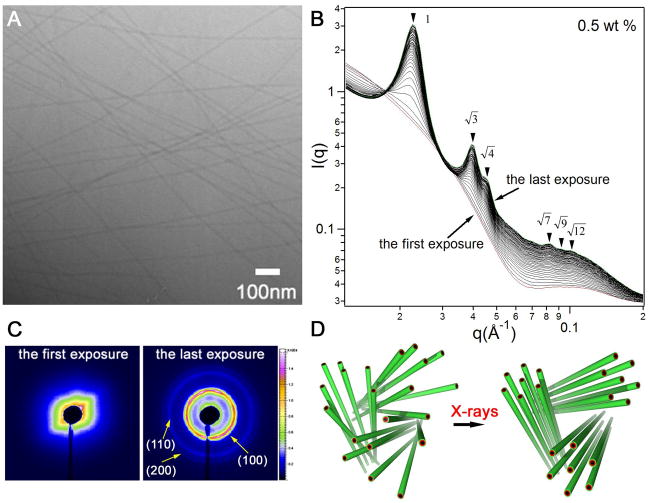
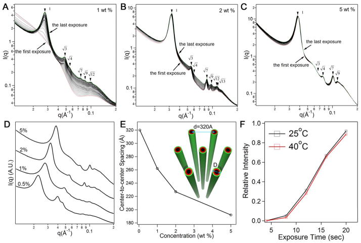
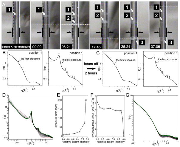
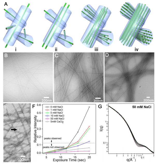
We report here crystallization at long range in networks of like-charge supramolecular peptide filaments mediated by repulsive forces. The crystallization is spontaneous beyond a given concentration of the molecules that form the filaments but can be triggered by x-rays at lower concentrations. The crystalline domains formed by x-ray irradiation, with interfilament separations of up to 320 angstroms, can be stable for hours after the beam is turned off, and ions that screen charges on the filaments suppress ordering. We hypothesize that the stability of crystalline domains emerges from a balance of repulsive tensions linked to native or x-ray-induced charges and the mechanical compressive entrapment of filaments within a network. Similar phenomena may occur naturally in the cytoskeleton of cells and, if induced externally in biological or artificial systems, lead to possible biomedical and lithographic functions.


Figures

Figure 1
Figure 1
X-ray irradiation triggered crystallization of self-assembling filaments at 0.5 wt % aqueous solutions of a peptide amphiphile. (A) Representative cryo-TEM image of filaments formed in 0.5 wt % solutions of peptide amphiphile; (B) a series of 50 consecutive scattering profiles of 0.5 wt % solutions obtained from the same sample area with four second exposure each (dose rate: 4806 Gy/second). The measurements were performed at room temperature with a monochromic X-rays at 15keV, and the typical incident X-ray flux on the sample was ~1×10¹² photons/s with a 0.2×0.3 mm2 collimator (samples were placed inside quartz capillaries of a diameter of 2 mm). The first and the last spectra are colored red and green, respectively; (C) 2D scattering patterns corresponding to the first and last exposures in B; (D) schematic representation of X-ray triggered organization of peptide filaments from a disordered state to a hexagonally ordered state.
Figure 2
Figure 2
Concentration dependence and temperature effect of X-ray triggered filament crystallization. (A) 50 consecutive scattering profiles from 1 wt % aqueous solutions (exposure time: 4 seconds); (B) 30 consecutive scattering profiles from 2 wt % solutions (exposure time: 2 seconds), and 5 wt % solutions (exposure time: 1 second) (C) (the dose rate for the experiments in (A–C) was 4806 Gy/second). The first and the last profiles in A, B, and C are colored red and green, respectively; (D) a replot of X-ray scans corresponding to the last exposure at concentrations of 0.5 wt %, 1 wt %, 2 wt % and 5 wt %; (E) a plot of center-to-center spacings among filaments as a function of peptide amphiphile concentration (the inset shows a schematic representation of the largest spacing observed among filaments in 0.5 wt % solutions); (F) effect of temperature on X-ray triggered ordering of the filaments in a 1 wt % solution (the relative intensity is given by (In−I4)/I4 where n denotes the accumulated exposure time and I4 is the intensity value at the location of the principal Bragg peak in the first scattering profile obtained after X-ray irradiation).
Figure 3
Figure 3
Reversible crystallization of filaments triggered by X-ray irradiation and the effects of the X-ray beam attenuation and photon energy. (A) A series of sequential photographs of the quartz capillary loaded with sample reveal the optical appearance changes before and after X-ray irradiation. The parallel black lines in the photographs demarcate the edges of the capillary and the white arrows point to the three spots (labeled as 1, 2, and 3) irradiated with X-rays for 200 s (the cylindrical shape visible within the space of the capillary is just an artifact of the photograph). From left to right, the first photograph corresponds to a sample-loaded capillary before X-ray irradiation and the second one shows opacity in spot 1 after 200 s irradiation (indicated as time 00:00 in the lower left hand corner of the photograph). Subsequent photographs from left to right reveal changes in the appearance of spots 1, 2 and 3 after the elapsed times indicated in the left hand corner of the photographs (minutes:seconds). The opacity in the irradiated spots dimmed eventually in the absence of the X-ray beam; (B) one-dimensional scattering profile after the first X-ray exposure of 4 seconds at position 1 and after the last of 49 additional exposures on the same position (all exposures lasted 4 s and the dose rate was 4806 Gy/second); (C) after a waiting period of two hours with the beam off, the experiment in (B) was repeated on position 1 and similar scattering profiles were obtained; (D) 50 consecutive scattering profiles from 1 wt % aqueous solution with a photon energy of 9 keV (exposure time = 2 seconds; photon flux = 2.2 × 10¹¹ photons/second; beam size = 0.37 mm × 0.31 mm; dose rate: 1037 Gy/second). The first and last spectra are colored red and green, respectively; (E) plot of accumulated X-ray exposure time required for observation of the principal Bragg peak as a function of relative beam intensity; (F) plot of the reciprocal of accumulated X-ray dose as a function of relative beam intensity; (G) the first 50 consecutive scattering profiles from 1 wt % aqueous solution (exposure time = 16 seconds; photon flux = 1.5 × 10¹⁰ photons/second; beam size = 0.37 mm × 0.31 mm; dose rate = 39.22 Gy/second).
Figure 4
Figure 4
(A) Schematic illustration of the proposed templating model for filament bundle formation during self-assembly. i, At early stages of self-assembly, interlocking of long filaments leads to the formation of a channel around each one of them. The long filament has many degrees of freedom within its channel, but cannot leave it or allow others enter its space. The volume of this imaginary channel should be determined mostly by the bulk density of long filaments; ii, newly formed long filaments from smaller ones and from monomer are confined to grow within these channels; iii, further growth causes the formation of bundles of aligned long filaments; iv, at high concentrations hexagonally ordered filaments are spontaneously formed as a result of repulsive electrostatic forces and network confinement; (B) cryo-TEM image of filament bundles in 0.5 wt % solutions, 1 wt % solutions (C), and 2 wt % solutions (D); (E) negatively stained TEM image of very early stages of self-assembly revealing clearly the presence of small aggregates (marked with black arrow) coexisting with loosely packed long filaments at 0.1 wt % (this image offers support for the templating mechanism for bundle formation explained in (A)); (F) the effect of added salts on X-ray triggered crystallization of filaments: plot of the intensity of the primary Bragg peak as a function of exposure time to X-rays in 1 wt % solutions, when the intensity plotted is below the dotted line the Bragg peak was not observed; (G) 50 X-ray scattering profiles corresponding to a 1 wt % solution containing 50 mM concentration of NaCl obtained sequentially after 50 exposures of 4 s each to the X-ray beam (dose rate: 4806 Gy/second).

Similar articles

Cited by

References

    1. Song WH, Kinloch IA, Windle AH. Science. 2003 Nov;302:1363. - PubMed
    1. Lee SW, Mao CB, Flynn CE, Belcher AM. Science. 2002 May;296:892. - PubMed
    1. Nakata M, et al. Science. 2007 Nov;318:1276. - PubMed
    1. Needleman DJ, et al. Proc Natl Acad Sci U S A. 2004 Nov;101:16099. - PMC - PubMed
    1. Pelletier O, et al. Phys Rev Lett. 2003 Oct;91:4. - PubMed

Publication types