. 2010 May;51(5):2372-80.
doi: 10.1167/iovs.09-4895. Epub 2009 Dec 17.

Visual Arrestin 1 contributes to cone photoreceptor survival and light adaptation

Affiliations

Visual Arrestin 1 contributes to cone photoreceptor survival and light adaptation

Bruce M Brown et al. Invest Ophthalmol Vis Sci. 2010 May.

Abstract

Purpose: To evaluate morphologic and functional contributions of Arrestin 1 (Arr1) and Arrestin 4 (Arr4) in cone photoreceptors, the authors examined the phenotypes of visual arrestin knockout mice (Arr1(-/-), Arr4(-/-), Arr1(-/-)Arr4(-/-) [Arr-DKO]) reared in darkness.

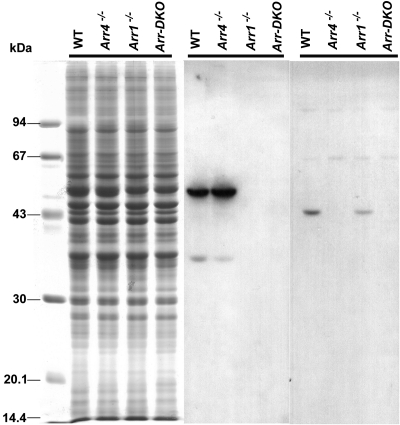
Methods: Retinal rods and cones were evaluated in wild-type (WT), Arr1(-/-), Arr4(-/-), and Arr-DKO mice using quantitative morphologic analysis, immunoblot, immunohistochemistry, TUNEL, and electroretinographic (ERG) techniques.

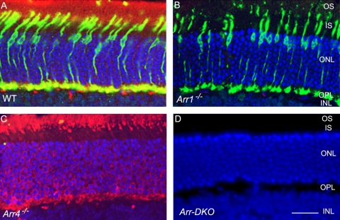
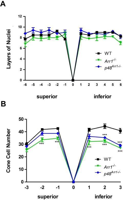
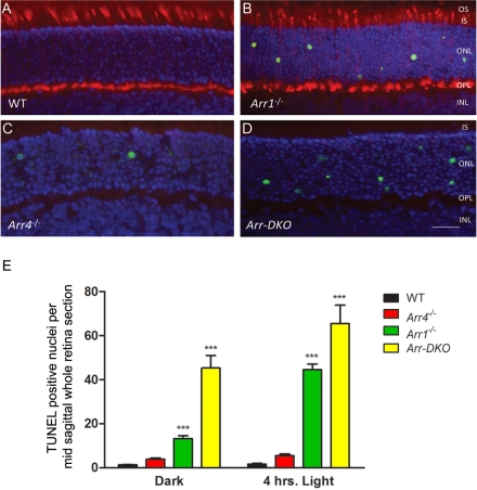
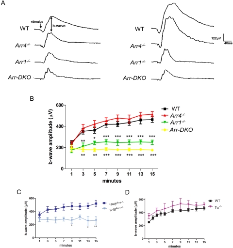
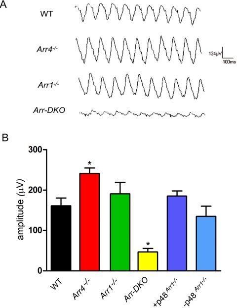
Results: Compared with either Arr4(-/-) or WT, Arr1(-/-) and Arr-DKO mice had increased apoptotic nuclei in their retinal outer nuclear layer (ONL) at postnatal day (P) 22. By P60, cone density was significantly diminished, but the ONL appeared normal. After 1 minute of background illumination, cone ERG b-wave amplitudes were similar in WT and all Arr KO mice. However, by 3 minutes and continuing through 15 minutes of light adaptation, the cone b-wave amplitudes of WT and Arr4(-/-) mice increased significantly over those of the Arr1(-/-) and Arr-DKO mice, which demonstrated no cone b-wave amplitude increase. In contrast, ERG flicker analysis after the 15-minute light adaptation period demonstrated no loss in amplitude for either Arr1(-/-) or Arr4(-/-) mice, whereas Arr-DKO had significantly lower amplitudes. When Arr1 expression was restored in Arr1(-/-) mice (+p48(Arr1-/-)), normal cone density and light-adapted ERG b-wave amplitudes were observed.

Conclusions: In the adult dark-reared Arr1(-/-) and Arr-DKO mice, viable cones diminish over time. Arr1 expression is essential for cone photoreceptor survival and light adaptation, whereas either Arr1 or Arr4 is necessary for maintaining normal flicker responses.


Figures

Figure 1.
Figure 1.
Immunoblot analysis at P40 of visual arrestin expression in mice retinas from control (WT) and visual arrestin knockouts (Arr1−/−, Arr4−/−, Arr-DKO). Each lane represents ∼50 μg retinal proteins from each genotype applied to 11.5% SDS-PAGE and either Coomassie Blue stained (left) or transferred to PVDF membrane and incubated with pAb C10C10 (Arr1) (middle) or pAb mCAR-LUMIj (Arr4) (right), followed by the appropriate HRP-conjugated secondary antibody and processed for ECL. Molecular weight markers (in kDa) are identified on the left.
Figure 2.
Figure 2.
IHC analysis at P60 of visual arrestin expression in mice retinas from control (WT) and visual arrestin knockouts (Arr1−/−, Arr4−/−, Arr-DKO). Retinal frozen sections of WT (A), Arr1−/− (B), Arr4−/−(C), and Arr-DKO (D) were processed with Arr1 (mAb D9F2) and Arr4 (pAb mCAR-LUMIj) antibodies, followed by the appropriate secondary antibodies. Immunoreactive Arr1 is red (Texas red) and Arr4 is green (fluorescein). Dual immunolocalization of Arr1 and Arr4 is yellow. Nuclei are stained blue with DAPI. IS, inner segment; OS, outer segment; ONL, outer nuclear layer; OPL, outer plexiform layer; INL, inner nuclear layer. Original magnification, 40×. Scale bar, 20 μm.
Figure 3.
Figure 3.
Quantitative spider plot analysis of mouse retinas at P60 from control (WT), Arr1−/−, or p48Arr1−/−. (A) The total number of layers of nuclei in the ONL of hematoxylin and eosin–stained retinal sections through the optic nerve (0) was measured at 12 locations around the retina, six each in the superior and inferior hemispheres. At least four mice of each genotype were counted. Two-way ANOVA with Bonferroni posttests compared Arr1−/− or +p48Arr1−/− transgenic mice with WT at each segment measured. (B) The number of cones in the photoreceptor layer of mCAR-LUMIj immunologically stained retinal sections through the optic nerve (0) was counted in six 290-μm segments, three each in the superior and inferior hemispheres 100 μm from the optic nerve. At least four retinas of each genotype were counted. Two-way ANOVA with Bonferroni posttests compared KO mice with WT in all segments (***P < 0.001).
Figure 4.
Figure 4.
Fluorometric TUNEL analysis of mice retinas at P22 from control (WT) and visual arrestin knockouts (Arr1−/−, Arr4−/−, Arr-DKO). Images illustrating cell apoptosis in the ONL from frozen retinal sections from P22. (A) WT, (B) Arr1−/−, (C) Arr4−/−, (D) Arr-DKO. Mice were killed in the dark and analyzed using the TUNEL system, with immunologic staining with Arr4 (pAb mCAR-LUMIj antibody) followed by the appropriate secondary antibody. Immunoreactive Arr4 is red (Texas red), and nuclei are stained blue with DAPI. Original magnification, 40×. Scale bar, 20 μm. (E) Histogram analysis summarizes these data from cell counting of all TUNEL-positive nuclei on averaging the total from three adjacent midsagittal whole retinal sections from at least four mice for each genotype killed either in the dark or after 4 hours of 1400 lux illumination. Bars represent ±SEM. Two-way ANOVA with Bonferroni posttests compared visual arrestin knockout mice with WT mice (***P < 0.001).
Figure 5.
Figure 5.
ERG analysis of P35 to P60 control (WT), visual arrestin knockouts (Arr1−/−, Arr4−/−, Arr-DKO), p48Arr−/−, and Tα−/− mice. A bifurcated glass fiber optic delivered both maximum intensity (2.01 log cd · s/m2) 10-μs bright flashes and a 200 cd/m2 steady background white light to a level 1 cm from the cornea. (A) Representative responses to bright flashes (stimulus) of WT and visual arrestin KO mice at 1 minute (left waveform data) and after 15 minutes of continuous background illumination (right waveform data). Average photopic b-wave amplitudes (μV) recorded every 2 minutes during 15 minutes of light adaptation of the (B) WT and visual arrestin knockout mice, (C) transgenic p48 mice bred onto an Arr1 null background (Arr1−/−) with confirmed Arr1 (+p48Arr1−/−) or negative littermate controls (−p48Arr1−/−), and (D) rod alpha transducin (Tα−/−) mice. Two-way ANOVA with Bonferroni posttests used for statistical comparisons with controls (***P < 0.001; **P < 0.01; *P < 0.05).
Figure 6.
Figure 6.
ERG Flicker analysis of control (WT), visual arrestin knockouts (Arr4−/−, Arr1−/−, Arr-DKO), +p48Arr1−/−, or −p48Arr1−/− mice. (A) Representative 10-Hz flicker responses of WT and visual arrestin knockout mice measured after 16 minutes of background illumination. (B) 10-Hz flicker responses of WT, Arr4−/−, Arr1−/−, Arr-DKO, and p48 transgenic mice (+p48Arr1−/−) and their negative littermate controls (−p48Arr1−/−). Two-way ANOVA with Bonferroni posttests compared transgenic mice with WT mice (*P < 0.05).

Similar articles

Cited by

References

    1. Wacker WB, Lipton MM. Experimental allergic uveitis: homologous retina as uveitogenic antigen. Nature 1965;206:253–254 - PubMed
    1. Wacker WB, Donoso LA, Kalsow CM, Yankeelov JA, Jr, Organisciak DT. Experimental allergic uveitis: isolation, characterization, and localization of a soluble uveitopathogenic antigen from bovine retina. J Immunol 1977;119:1949–1958 - PubMed
    1. Kuhn H. Light-regulated binding of rhodopsin kinase and other proteins to cattle photoreceptor membranes. Biochemistry 1978;17:4389–4395 - PubMed
    1. Pfister C, Chabre M, Plouet J, et al. Retinal S antigen identified as the 48K protein regulating light-dependent phosphodiesterase in rods. Science 1985;228:891–893 - PubMed
    1. Xu J, Dodd RL, Makino CL, Simon MI, Baylor DA, Chen J. Prolonged photoresponses in transgenic mouse rods lacking arrestin. Nature 1997;389:505–509 - PubMed

Publication types