Differential distribution of blood and lymphatic vessels in the murine cornea
- PMID: 20019372
- PMCID: PMC2868485
- DOI: 10.1167/iovs.09-4505
Differential distribution of blood and lymphatic vessels in the murine cornea
Abstract
Purpose: Because of its unique characteristics, the cornea has been widely used for blood and lymphatic vessel research. However, whether limbal or corneal vessels are evenly distributed under normal or inflamed conditions has never been studied. The purpose of this study was to investigate this question and to examine whether and how the distribution patterns change during corneal inflammatory lymphangiogenesis (LG) and hemangiogenesis (HG).
Methods: Corneal inflammatory LG and HG were induced in two most commonly used mouse strains, BALB/c and C57BL/6 (6-8 weeks of age), by a standardized two-suture placement model. Oriented flat-mount corneas together with the limbal tissues were used for immunofluorescence microscope studies. Blood and lymphatic vessels under normal and inflamed conditions were analyzed and quantified to compare their distributions.
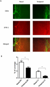
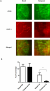
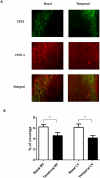
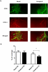
Results: The data demonstrate, for the first time, greater distribution of both blood and lymphatic vessels in the nasal side in normal murine limbal areas. This nasal-dominant pattern was maintained during corneal inflammatory LG, whereas it was lost for HG.
Conclusions: Blood and lymphatic vessels are not evenly distributed in normal limbal areas. Furthermore, corneal LG and HG respond differently to inflammatory stimuli. These new findings will shed some light on corneal physiology and pathogenesis and on the development of experimental models and therapeutic strategies for corneal diseases.
Figures

Similar articles
-
Time course of angiogenesis and lymphangiogenesis after brief corneal inflammation.Cornea. 2006 May;25(4):443-7. doi: 10.1097/01.ico.0000183485.85636.ff. Cornea. 2006. PMID: 16670483
-
Short- and long-term corneal vascular effects of tafluprost eye drops.Graefes Arch Clin Exp Ophthalmol. 2013 Aug;251(8):1919-27. doi: 10.1007/s00417-013-2345-0. Epub 2013 Apr 28. Graefes Arch Clin Exp Ophthalmol. 2013. PMID: 23624591
-
Kinetics of Angiogenic Responses in Corneal Transplantation.Cornea. 2017 Apr;36(4):491-496. doi: 10.1097/ICO.0000000000001127. Cornea. 2017. PMID: 28060028 Free PMC article.
-
Corneal lymphangiogenesis: implications in immunity.Semin Ophthalmol. 2009 May-Jun;24(3):135-8. doi: 10.1080/08820530902801320. Semin Ophthalmol. 2009. PMID: 19437348 Review.
-
Lymphangiogenesis Guidance Mechanisms and Therapeutic Implications in Pathological States of the Cornea.Cells. 2023 Jan 14;12(2):319. doi: 10.3390/cells12020319. Cells. 2023. PMID: 36672254 Free PMC article. Review.
Cited by
-
Very late antigen-1 mediates corneal lymphangiogenesis.Invest Ophthalmol Vis Sci. 2011 Jul 1;52(7):4808-12. doi: 10.1167/iovs.10-6580. Invest Ophthalmol Vis Sci. 2011. PMID: 21372020 Free PMC article.
-
Persistence of reduced expression of putative stem cell markers and slow wound healing in cultured diabetic limbal epithelial cells.Mol Vis. 2015 Dec 30;21:1357-67. eCollection 2015. Mol Vis. 2015. PMID: 26788028 Free PMC article.
-
Lymphatics in Eye Fluid Homeostasis: Minor Contributors or Significant Actors?Biology (Basel). 2021 Jun 25;10(7):582. doi: 10.3390/biology10070582. Biology (Basel). 2021. PMID: 34201989 Free PMC article. Review.
-
A detailed survey of the murine limbus, its stem cell distribution, and its boundaries with the cornea and conjunctiva.Stem Cells Transl Med. 2024 Oct 10;13(10):1015-1027. doi: 10.1093/stcltm/szae055. Stem Cells Transl Med. 2024. PMID: 39077915 Free PMC article.
-
The Role of the miR-21/SPRY2 Axis in Modulating Proangiogenic Factors, Epithelial Phenotypes, and Wound Healing in Corneal Epithelial Cells.Invest Ophthalmol Vis Sci. 2019 Sep 3;60(12):3854-3862. doi: 10.1167/iovs.19-27013. Invest Ophthalmol Vis Sci. 2019. PMID: 31529118 Free PMC article.
References
-
- Brown P. Lymphatic system: unlocking the drains. Nature 2005;436:456–458 - PubMed
-
- Jackson DG. Biology of the lymphatic marker LYVE-1 and applications in research into lymphatic trafficking and lymphangiogenesis. APMIS 2004;112:526–538 - PubMed
-
- Chang L, Kaipainen A, Folkman J. Lymphangiogenesis new mechanisms. Ann N Y Acad Sci 2002;979:111–119 - PubMed
-
- Oliver G, Detmar M. The rediscovery of the lymphatic system: old and new insights into the development and biological function of the lymphatic vasculature. Genes Dev 2002;16:773–783 - PubMed
-
- Alitalo K, Tammela T, Petrova TV. Lymphangiogenesis in development and human disease. Nature 2005;438:946–953 - PubMed
Publication types
MeSH terms
Substances
LinkOut - more resources
Full Text Sources
Other Literature Sources

