Clinical validation study of percutaneous cochlear access using patient-customized microstereotactic frames
- PMID: 20019561
- PMCID: PMC2845321
- DOI: 10.1097/MAO.0b013e3181c2f81a
Clinical validation study of percutaneous cochlear access using patient-customized microstereotactic frames
Abstract
Objective: Percutaneous cochlear implant (PCI) surgery consists of drilling a single trough from the lateral cranium to the cochlea avoiding vital anatomy. To accomplish PCI, we use a patient-customized microstereotactic frame, which we call a "microtable" because it consists of a small tabletop sitting on legs. The orientation of the legs controls the alignment of the tabletop such that it is perpendicular to a specified trajectory.
Study design: Prospective.
Setting: Tertiary referral center.
Patients: Thirteen patients (18 ears) undergoing traditional cochlear implant surgery.
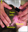
Interventions: With institutional review board approval, each patient had 3 fiducial markers implanted in bone surrounding the ear. Temporal bone computed tomographic scans were obtained, and the markers were localized, as was vital anatomy. A linear trajectory from the lateral cranium through the facial recess to the cochlea was planned. A microtable was fabricated to follow the specified trajectory.
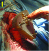
Main outcome measures: After mastoidectomy and posterior tympanotomy, accuracy of trajectories was validated by mounting the microtables on the bone-implanted markers and then passing sham drill bits across the facial recess to the cochlea. The distance from the drill to vital anatomy was measured.
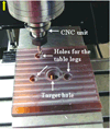
Results: Microtables were constructed on a computer-numeric-control milling machine in less than 5 minutes each. Successful access across the facial recess to the cochlea was achieved in all 18 cases. The mean +/- SD distance was 1.20 +/- 0.36 mm from midportion of the drill to the facial nerve and 1.25 +/- 0.33 mm from the chorda tympani.
Conclusion: These results demonstrate the feasibility of PCI access using customized microstereotactic frames.
Figures


References
-
- Warren FM, Balachandran R, Fitzpatrick JM, Labadie RF. Percutaneous Cochlear Access Using Bone-Mounted, Customized Drill Guides: Demonstration of Concept In-Vitro. Otology & Neurotology. 2007;28(3):325–329. - PubMed
-
- Balachandran R, Mitchell J, Dawant B, Fitzpatrick JM. Evaluation of Targeting Frames for Deep-Brain Stimulation Using Virtual Targets. Presented at the Proc. IEEE Intl. Symp. On Biomedical Imaging 2007; Arlington, VA. 2007. Apr, pp. 1184–1187.
Publication types
MeSH terms
Grants and funding
LinkOut - more resources
Full Text Sources
Other Literature Sources
Miscellaneous

