. 2010 Jan 20;29(2):410-23.
doi: 10.1038/emboj.2009.357. Epub 2009 Dec 17.

DNA polymerase beta is critical for mouse meiotic synapsis

Affiliations

DNA polymerase beta is critical for mouse meiotic synapsis

Dawit Kidane et al. EMBO J. .

Abstract

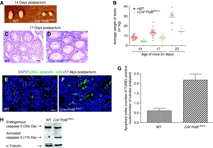
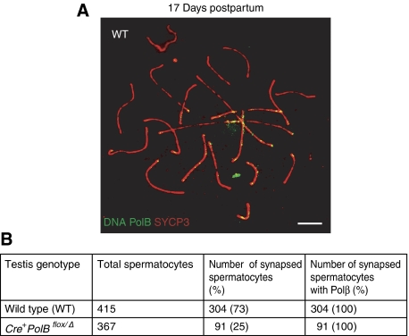
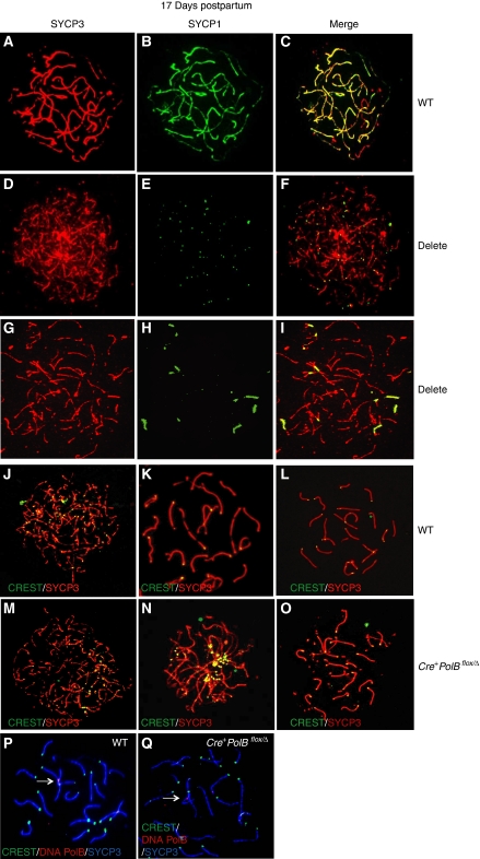
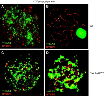
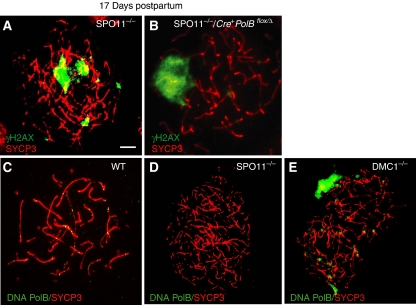
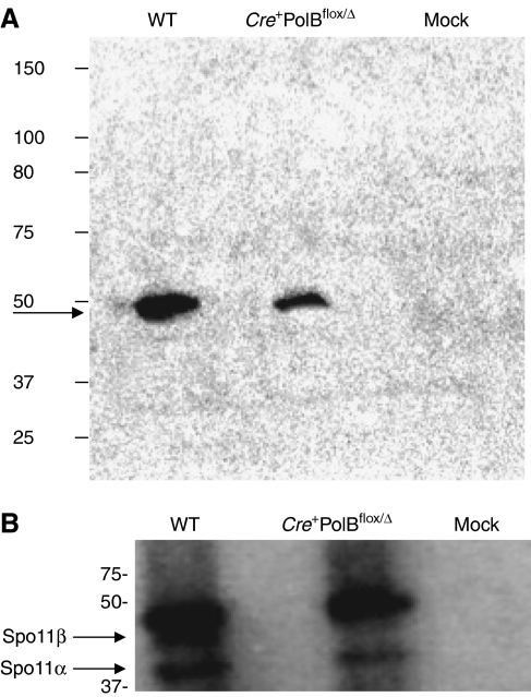
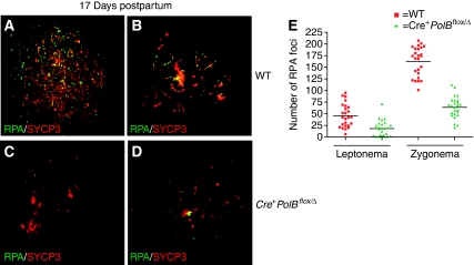
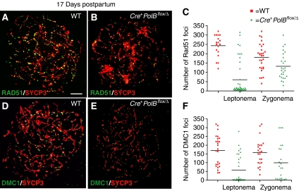
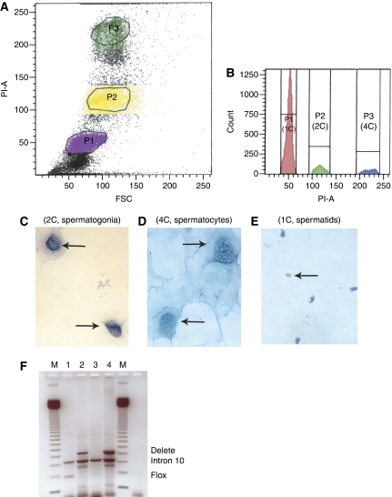
We have shown earlier that DNA polymerase beta (Pol beta) localizes to the synaptonemal complex (SC) during Prophase I of meiosis in mice. Pol beta localizes to synapsed axes during zygonema and pachynema, and it associates with the ends of bivalents during late pachynema and diplonema. To test whether these localization patterns reflect a function for Pol beta in recombination and/or synapsis, we used conditional gene targeting to delete the PolB gene from germ cells. We find that Pol beta-deficient spermatocytes are defective in meiotic chromosome synapsis and undergo apoptosis during Prophase I. We also find that SPO11-dependent gammaH2AX persists on meiotic chromatin, indicating that Pol beta is critical for the repair of SPO11-induced double-strand breaks (DSBs). Pol beta-deficient spermatocytes yielded reduced steady-state levels of the SPO11-oligonucleotide complexes that are formed when SPO11 is removed from the ends of DSBs, and cytological experiments revealed that chromosome-associated foci of replication protein A (RPA), RAD51 and DMC1 are less abundant in Pol beta-deficient spermatocyte nuclei. Localization of Pol beta to meiotic chromosomes requires the formation of SPO11-dependent DSBs. Taken together, these findings strongly indicate that Pol beta is required at a very early step in the processing of meiotic DSBs, at or before the removal of SPO11 from DSB ends and the generation of the 3' single-stranded tails necessary for subsequent strand exchange. The chromosome synapsis defects and Prophase I apoptosis of Pol beta-deficient spermatocytes are likely a direct consequence of these recombination defects.


Conflict of interest statement

The authors declare that they have no conflict of interest.

Figures

Figure 1
Figure 1
Molecular characterization of PolB mice. (A) Schematic representation of the PolB gene locus before and after exposure to Cre. The WT, flox and delete alleles are pictured. The loxP sites in the floxed allele are denoted by filled arrowheads. When crossed with mice expressing Cre protein from the TNAP promoter, there is site-specific recombination between the loxP sites, leading to deletion of part of the promoter region (denoted by a bent arrow), and exon 1. This leaves one loxP site in the deleted allele. The flox allele is detected by amplification with the βmetF1 and βmetF2 primers, shown above the floxed allele in the cartoon. The deleted allele is detected by amplification with the βF1 and βR1 primers shown above and below, respectively, the deleted allele in the cartoon. (B) The PolBflox and PolBdelete alleles can be detected by PCR. Genomic DNA was isolated from tail clippings and used as templates in a PCR reaction with the primers as described in Materials and methods. An aliquot of the PCR reaction was resolved on an agarose gel, and the image was captured using a Multimage Light Camera (Alpha Innotech). M, marker (base pairs); lane 1, tail DNA with the flox allele alone; lane 2, DNA with WT allele alone; lane 3, DNA with both WT and flox alleles; lane 4, DNA with the delete allele; lane 5, DNA with the Cre+ transgene. (C) The PolB deletion index can be estimated by Southern blotting. Genomic DNA was isolated from testes, digested with BamHI, resolved on an agarose gel, and analysed by Southern blotting as described in Materials and methods. Lane 1, mouse DNA carrying only the WT allele; lane 2, mouse DNA carrying only the PolBflox allele; lane 3, Cre mouse carrying both PolBflox and PolBdelete alleles; lane 4, Cre+ mouse carrying both the PolBflox and PolBdelete alleles, present at 10 and 90% of total, respectively, as determined by image analysis. The presence of the Cre+ transgene was determined by PCR as in (B).
Figure 2
Figure 2
Pol β-deficient seminferous tubules have few germ cells. (A) Testes of 14-day-old Cre+PolBflox/Δ mice (right) are smaller than those of WT (left). (B) Average testis weight (in mg) versus age (▪: WT mice (red); ▴: Cre+PolBflox/Δ mice (green)). (C) Tubule histology of 17 WT pups. (D) Histology of tubules of 17-day-old Cre+ PolBflox/Δ pups. Image was taken at 400 × magnification (scale bar=10 μM). Note that the pups with the Cre+ transgene exhibit aberrant testis morphology. Black and red symbols designated by (*) denote tubules with few and moderate numbers of cells, respectively. (E, F) TUNEL assay of two seminiferous tubules (tubules marked 1 and 2) from 17-day-old WT and Cre+PolBflox/Δ mice. Image taken at 200 × magnification (scale bar=5 μM). (G) Quantification of TUNEL assay. (H) Activation of caspase-3 in Cre+PolBflox/Δ spermatocytes. Western blot analysis was performed using whole testis extracts from 17-day-old mice and immunoblotted with anti-caspase-3 antibody. 1, WT; 2, Cre+PolBflox/Δ mice.
Figure 3
Figure 3
Pol β-deficient germ cells do not progress through meiosis. (A) Example of a spermatocyte nucleus in the pachytene substage of meiotic Prophase I from a 17-day-old WT mouse. The bivalents are stained with SYCP3 (red) and Pol β (green). (B) Tabulation of cells exhibiting extensive synapsis in each of two genetic backgrounds. Genotype obtained by PCR analysis of tail DNA. Staging of randomly chosen nuclei was assessed by the staining pattern of SYCP3 antiserum and the DAPI image. At least three different 17-day-old mice of each genotype were assessed for progression to pachynema.
Figure 4
Figure 4
Pol β is critical for synapsis. (AC) Example of spermatocyte nuclei from a 17-day-old WT pup. (A) SYCP3 (red); (B) SYCP1 (green); (C) merge of panels (A) and (B). (DI) Examples of spermatocyte nuclei from a 17-day-old Cre+PolBflox/Δ pup. (D, G) SYCP3 (red); (E, H) SYCP1 (green); (F, I) merge of panels (D) and (E) and (G) and (H), respectively. (JL) Example of centromere localization (CREST) in WT spermatocyte nuclei. (J) Zygotene substage stained with CREST (n=40); (K) early pachynema (n=21); (L) pachynema. (MO) Example of CREST localization from Cre+PolBflox/Δ spermatocyte nuclei. (M) Zygonema, (n=40); (N) late zygonema (n=40); (O) pachynema like (n=29). SYCP3 (red), CREST (green). (PQ) Examples of 17-day-old nuclei labelled with CREST (green), SYCP3 (white) and Pol β (red). (P) WT nucleus. (Q, P) Nucleus from Cre+PolBflox/Δ pup that appears to have progressed to pachynema. White arrows point to Pol β foci.
Figure 5
Figure 5
Meiotic DSBs are generated but not repaired in Pol β-deficient spermatocytes. Shown are surface spreads of spermatocyte nuclei from WT (A, B) and Cre+PolBflox/Δ (C, D) 17-day-old mice. Each of the nuclei were stained with antisera raised against γH2AX (green) and SYCP3 (red). Note that this signal is much more intense and persists in the nuclei isolated from Cre+PolBflox/Δ pups, whereas it only stains the sex body in WT pups and those without the Cre transgene.
Figure 6
Figure 6
Pol β functions in the repair of SPO11-induced double-strand breaks. The PolBflox/Δ mice with or without the Cre transgene were crossed into a SPO11 background. (A) Example of a spermatocyte nucleus from 17-day-old SPO11−/− mice. The axial elements are stained with SYCP3 (red) and γH2AX (green). (B) Example of a spermatocyte nucleus from Cre+PolBflox/Δ SPO11−/− mice. Note the presence of a pseudo sex body and other staining by γH2AX on the autosomes. (CE) Examples of localization of Pol β on spermatocyte spreads obtained from (C) WT; (D) SPO11−/−; (E) DMC1−/− (SYCP3 (red), Pol β (green)). Note that there is more than one nucleus in each panel.
Figure 7
Figure 7
SPO11 is not completely released from the SCs of Pol β-deficient spermatocytes. (A) Whole testis extracts, isolated from 17-day-old mice, were incubated with SPO11 antiserum for 2 h, and then with agarose beads overnight. After centrifugation and washing, as described in Materials and methods, TdT and γ-32P were added to label the SPO11-DNA complex. Lane 1, extracts from WT spermatocytes; lane 2, extracts from Cre+PolBflox/Δ; lane 3, mock control (background). In this case, PBS was added to the extracts prepared from WT spermatocytes instead of antiserum against SPO11. Arrow points to SPO11 DNA complex. See Materials and methods for details. (B) Immunoblotting of the blot in (A) with monoclonal antibody raised against SPO11. The secondary antibody in this case with mouse HRP-protein A to reduce background. The bands corresponding to SPO11α and SPO11β are denoted with arrows and labels.
Figure 8
Figure 8
Fewer RPA foci associate with the SC in Pol β-deficient spermatocytes. Spread spermatocyte nuclei of 17-day-old WT (top two panels; A, B) and Cre+PolBflox/Δ mice (bottom two panels; C, D) during leptonema (left) and zygonema (right). The SC is visualized with antiserum raised against SYCP3 (red) and RPA is visualized with antiserum raised against the 34-kDa subunit of RPA (green). Note that RPA appears to be associated with the synapsed axes during zygonema. (E) Quantification of the number of foci per nucleus of RPA in leptonema and pachynema in the WT and Pol β-deficient spermatocytes.
Figure 9
Figure 9
Fewer RAD51 and DMC1 foci are observed on the SCs of Pol β-deficient spermatocytes. (A, B) Examples of RAD51 localization in 17-day-old mice; (A) WT; (B) Cre+PolBflox/Δ; (C) Scatter plot of leptotene/zygotene spermatocyte nuclei that display RAD51 foci versus the number of foci per nucleus. Red squares indicate focus counts in WT cells; green triangles indicate counts in mutant cells. RAD51 focus counts in leptotene cells (mean±s.e.m.) were 242±13 in WT and 60±14 in spermatocytes from Cre+PolBflox/Δ mice (P<0.0001). Counts in zygotene cells were 180±13 and 132±13 for WT and mutant, respectively (P<0.01). (DF) Examples of DMC1 localization; (D) WT; (E) Cre+PolBflox/Δ; (F), Scatter plot of leptotene/zygotene spermatocyte nuclei that display DMC1 foci versus the number of foci per nucleus. DMC1 focus counts in leptotene cells were 169±15 and 58±14 in WT and mutant, respectively (P<0.0001). Counts in zygotene cells were 159±15 and 99±21, respectively (P<0.01). Note that P-value was calculated by using Mann–Whitney test.
Figure 10
Figure 10
PolB undergoes excision before cells reach the spermatid stage. (A, B) Display FACS analysis of testicular cells from 10-week-old Cre+PolBflox/Δ mice. FSC is forward scatter and PI-A is propidium iodide staining. Each population was examined using light microscopy as shown in panels (CE), which contain spermatogonia (arrows), spermatocytes (arrows) and spermatids (mostly elongating and arrow points to a round spermatid), respectively. (F) Shows an example of PCR amplification of DNA isolated from various testicular cell fractions from Cre+PolBflox/Δ mice. M is the marker. Lane 1, spermatocyte DNA with primers for Intron 10 and the flox allele; lane 2, spermatocyte DNA with primers for Intron 10 and the delete allele. The band that resolves just under the Intron 10 band in this and other lanes is unknown, but consistently appears when primers for Intron 10 and the delete allele are used in the PCR reaction; lane 3, spermatid DNA with primers for Intron 10 and the flox allele; lane 4, spermatid DNA with primers for Intron 10 and the delete allele.

Similar articles

Cited by

References

    1. Alcivar AA, Hake LE, Hecht NB (1992) DNA polymerase-beta and poly(ADP)ribose polymerase mRNAs are differentially expressed during the development of male germinal cells. Biol Reprod 46: 201–207 - PubMed
    1. Ashley T (2004) The mouse ‘tool box' for meiotic studies. Cytogenet Genome Res 105: 166–171 - PubMed
    1. Ashley T, Plug A (1998) Caught in the act: deducing meiotic function from protein immunolocalization. Curr Top Dev Biol 37: 201–239 - PubMed
    1. Ashley T, Plug AW, Xu J, Solari AJ, Reddy G, Golub EI, Ward DC (1995) Dynamic changes in Rad51 distribution on chromatin during meiosis in male and female vertebrates. Chromosoma 104: 19–28 - PubMed
    1. Bannister LA, Schimenti JC (2004) Homologous recombinational repair proteins in mouse meiosis. Cytogenet Genome Res 107: 191–200 - PubMed

Publication types