

Monoallelic but not biallelic loss of Dicer1 promotes tumorigenesis in vivo
- PMID: 20019750
- PMCID: PMC2892162
- DOI: 10.1038/cdd.2009.202
Monoallelic but not biallelic loss of Dicer1 promotes tumorigenesis in vivo
Abstract
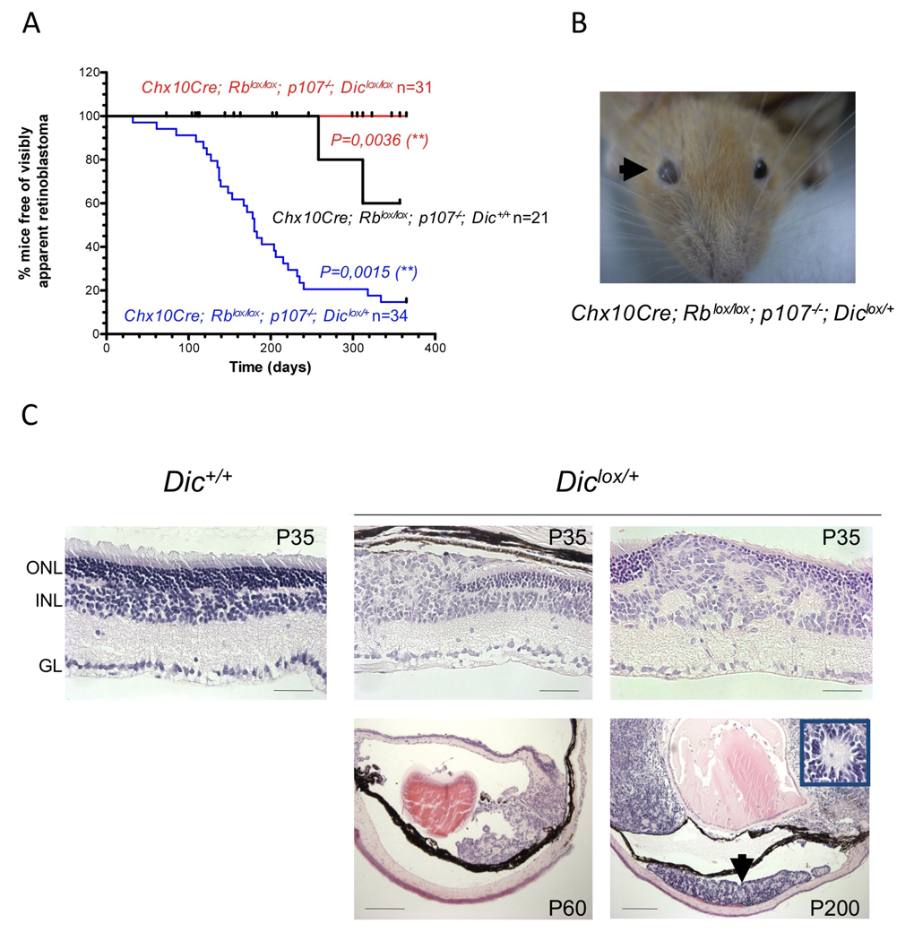
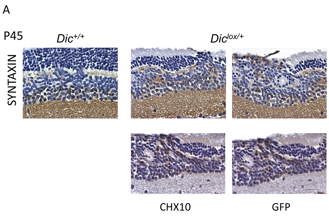
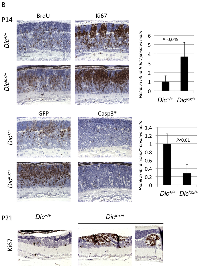
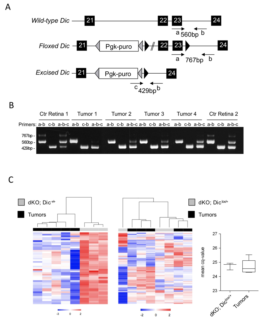
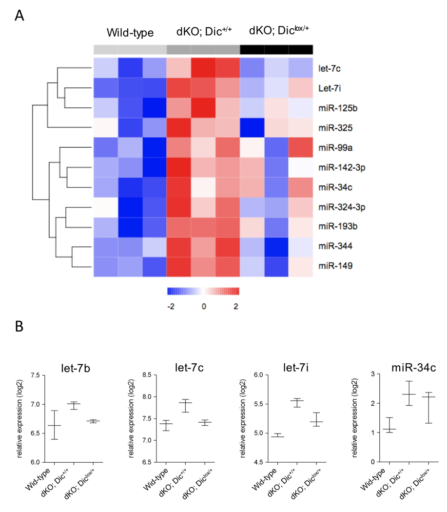
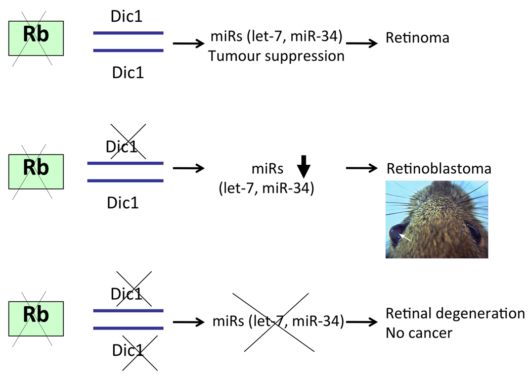
Human tumors are characterized by widespread reduction in microRNA (miRNA) expression, although it is unclear how such changes come about and whether they have an etiological role in the disease. Importantly, miRNA knockdown has been shown to enhance the tumorigenic potential of human lung adenocarcinoma cells. A defect in miRNA processing is one possible mechanism for global downregulation. To explore this possibility in more detail in vivo, we have manipulated Dicer1 gene dosage in a mouse model of retinoblastoma. We show that although monoallelic loss of Dicer1 does not affect normal retinal development, it dramatically accelerates tumor formation on a retinoblastoma-sensitized background. Importantly, these tumors retain one wild-type Dicer1 allele and exhibit only a partial decrease in miRNA processing. Accordingly, in silico analysis of human cancer genome data reveals frequent hemizygous, but not homozygous, deletions of DICER1. Strikingly, complete loss of Dicer1 function in mice did not accelerate retinoblastoma formation. miRNA profiling of these tumors identified members of the let-7 and miR-34 families as candidate tumor suppressors in retinoblastoma. We conclude that Dicer1 functions as a haploinsufficient tumor suppressor. This finding has implications for cancer etiology and cancer therapy.
Keywords: Dicer; haploinsufficiency; microRNA; retinoblastoma; tumor suppressor.
Conflict of interest statement
The authors declare no conflict of interest
Figures
Similar articles
-
Synthetic lethality between Rb, p53 and Dicer or miR-17-92 in retinal progenitors suppresses retinoblastoma formation.Nat Cell Biol. 2012 Sep;14(9):958-65. doi: 10.1038/ncb2556. Epub 2012 Aug 5. Nat Cell Biol. 2012. PMID: 22864477
-
Unique haploinsufficient role of the microRNA-processing molecule Dicer1 in a murine colitis-associated tumorigenesis model.PLoS One. 2013 Sep 2;8(9):e71969. doi: 10.1371/journal.pone.0071969. eCollection 2013. PLoS One. 2013. PMID: 24023722 Free PMC article.
-
Dicer1 functions as a haploinsufficient tumor suppressor.Genes Dev. 2009 Dec 1;23(23):2700-4. doi: 10.1101/gad.1848209. Epub 2009 Nov 10. Genes Dev. 2009. PMID: 19903759 Free PMC article.
-
The RB protein family in retinal development and retinoblastoma: new insights from new mouse models.Dev Neurosci. 2004;26(5-6):417-34. doi: 10.1159/000082284. Dev Neurosci. 2004. PMID: 15855771 Review.
-
Drosha: a new tumor suppressor in pineoblastoma.Genes Dev. 2025 Jun 2;39(11-12):677-678. doi: 10.1101/gad.352932.125. Genes Dev. 2025. PMID: 40345854 Free PMC article. Review.
Cited by
-
Downregulation of MST4 Underlies a Novel Inhibitory Role of MicroRNA Let-7a in the Progression of Retinoblastoma.Invest Ophthalmol Vis Sci. 2020 Jun 3;61(6):28. doi: 10.1167/iovs.61.6.28. Invest Ophthalmol Vis Sci. 2020. PMID: 32539131 Free PMC article.
-
Analyzing the Role of DICER1 Germline Variations in Papillary Thyroid Carcinoma.Eur Thyroid J. 2021 Feb;9(6):296-303. doi: 10.1159/000509183. Epub 2020 Aug 6. Eur Thyroid J. 2021. PMID: 33718253 Free PMC article.
-
microRNAs: New-Age Panacea in Cancer Therapeutics.Indian J Surg Oncol. 2021 Apr;12(Suppl 1):52-56. doi: 10.1007/s13193-020-01110-w. Epub 2020 Jun 6. Indian J Surg Oncol. 2021. PMID: 33994728 Free PMC article. Review.
-
The Clinical Significance of MicroRNAs in Colorectal Cancer Signaling Pathways: A Review.Glob Med Genet. 2023 Nov 22;10(4):315-323. doi: 10.1055/s-0043-1777094. eCollection 2023 Dec. Glob Med Genet. 2023. PMID: 38025193 Free PMC article. Review.
-
MicroRNAs shape circadian hepatic gene expression on a transcriptome-wide scale.Elife. 2014 May 27;3:e02510. doi: 10.7554/eLife.02510. Elife. 2014. PMID: 24867642 Free PMC article.
References
-
- Lu J, Getz G, Miska EA, Alvarez-Saavedra E, Lamb J, Peck D, Sweet-Cordero A, Ebert BL, Mak RH, Ferrando AA, et al. MicroRNA expression profiles classify human cancers. Nature. 2005;435:834–838. - PubMed
-
- Kumar MS, Lu J, Mercer KL, Golub TR, Jacks T. Impaired microRNA processing enhances cellular transformation and tumorigenesis. Nat Genet. 2007;39:673–677. - PubMed
Publication types
MeSH terms
Substances
Grants and funding
LinkOut - more resources
Full Text Sources
Other Literature Sources
Molecular Biology Databases
