. 2010 Feb;11(2):133-8.
doi: 10.1038/embor.2009.258. Epub 2009 Dec 18.

Mitochondrial dynamics regulate the RIG-I-like receptor antiviral pathway

Affiliations

Mitochondrial dynamics regulate the RIG-I-like receptor antiviral pathway

Céline Castanier et al. EMBO Rep. 2010 Feb.

Abstract

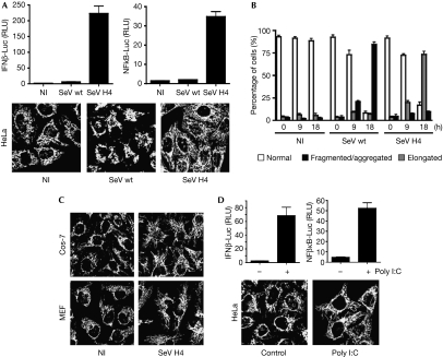
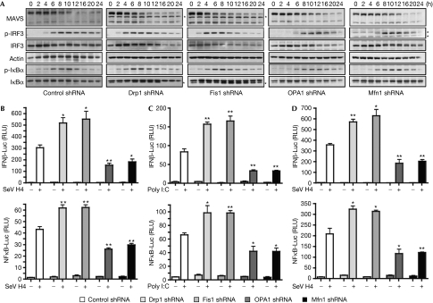
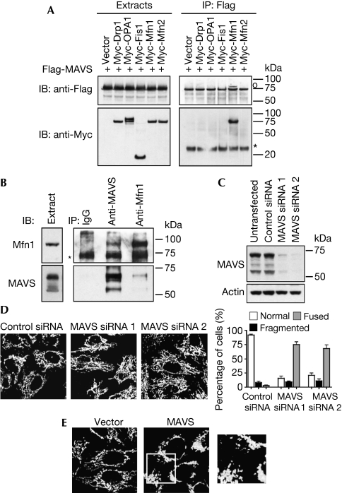
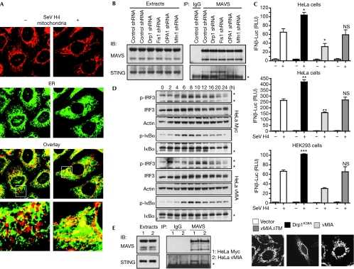
The intracellular retinoic acid-inducible gene I-like receptors (RLRs) sense viral ribonucleic acid and signal through the mitochondrial protein mitochondrial antiviral signalling (MAVS) to trigger the production of type I interferons and proinflammatory cytokines. In this study, we report that RLR activation promotes elongation of the mitochondrial network. Mimicking this elongation enhances signalling downstream from MAVS and favours the binding of MAVS to stimulator of interferon genes, an endoplasmic reticulum (ER) protein involved in the RLR pathway. By contrast, enforced mitochondrial fragmentation dampens signalling and reduces the association between both proteins. Our finding that MAVS is associated with a pool of mitofusin 1, a protein of the mitochondrial fusion machinery, suggests that MAVS is capable of regulating mitochondrial dynamics to facilitate the mitochondria-ER association required for signal transduction. Importantly, we observed that viral mitochondria-localized inhibitor of apoptosis, a cytomegalovirus (CMV) antiapoptotic protein that promotes mitochondrial fragmentation, inhibits signalling downstream from MAVS, suggesting a possible new immune modulation strategy of the CMV.


Conflict of interest statement

The authors declare that they have no conflict of interest.

Figures

Figure 1
Figure 1
RLR activation promotes elongation of the mitochondrial network. (A) HeLa cells were transfected with either an IFNβ promoter reporter or an NF-κB reporter, as well as with Renilla luciferase as an internal control. At 20 h after transfection cells were infected with SeV wt or SeV H4, or left uninfected. Luciferase assays were performed 9 h after infection and were normalized using Renilla luciferase activity. The error bars represent the standard deviation (s.d.) from the mean value obtained from three experiments. In parallel, mitochondrial morphology was assessed by immunofluorescence 18 h after infection. (B) Histogram for quantitating mitochondrial morphology in HeLa cells that were left either uninfected or infected with SeV wt or SeV H4 for 9 h or 18 h. Data are the means±s.d. of three independent experiments, with 300 cells per condition. (C) Cos-7 cells or MEFs were infected with SeV H4 for 18 h and mitochondrial morphology was observed by immunofluorescence. (D) HeLa cells were transfected as in panel (A). Twenty hours later cells were transfected with poly I:C (1 μg/ml) for 8 h and luciferase assays were performed. In parallel, mitochondrial morphology was assessed by immunofluorescence. IFN, interferon; Luc, luciferase; MEF, mouse embryonic fibroblast; NI, uninfected; NF-κB, nuclear factor-κB; RLR, RIG-I-like receptor; RLU, relative luciferase unit; SeV, Sendai virus; wt, wild type.
Figure 2
Figure 2
The RLR signalling pathway is modulated by alterations in mitochondrial dynamics. (A) HeLa cells were transfected with control, Drp1, Fis1, OPA1 or Mfn1 shRNA. After the selection of transfectants, cells were infected with SeV H4 and at various times after infection MAVS, p-IRF3, IRF3, p-IκBα and IκBα were analysed in cell extracts by immunoblotting. Actin was used as a protein loading control. *A probable non-specific protein band. Data shown are representative of three independent experiments. (B,C) IFNβ-Luc or NF-κB-Luc reporter plasmids were transfected into control cells or Drp1-, Fis1-, OPA1- or Mfn1-depleted cells, which were then infected with SeV H4 for 9 h (B) or transfected with poly I:C for 8 h (C), and then IFNβ induction and NF-κB activation were assessed. (D) The same conditions as in panel B, but HeLa cells were replaced by human embryonic kidney 293 cells. **0.001<P<0.01, *0.01<P<0.05. Drp1, dynamin-related protein 1; IFN, interferon; IRF, IFN regulatory factor; Luc, luciferase; MAVS, mitochondrial antiviral signalling; Mfn1, mitofusion 1; NF-κB, nuclear factor-κB; OPA1, optic atrophy type 1; RLR, RIG-I-like receptor; RLU, relative luciferase unit; SeV, Sendai virus; shRNA, short-hairpin RNA.
Figure 3
Figure 3
MAVS binds to Mfn1 and regulates mitochondrial morphology. (A) Human embryonic kidney 293 cells were transfected with Flag-MAVS and Myc constructs, where indicated, and cell extracts and immunoprecipitates (IP) were analysed by immunoblotting (IB). *IgG light chain; °a band from a previous immunoblot. (B) Endogenous Mfn1 and MAVS were immunoprecipitated from cell extracts with specific antibodies, and the presence of both proteins in immune complexes was examined by immunoblotting. *IgG heavy chain. (C,D) Control siRNA or siRNAs targeting MAVS were transfected into HeLa cells. Knockdown of MAVS was confirmed by immunoblotting (C) and mitochondrial morphology was analysed by immunofluorescence (D). (E) Control vector or MAVS-expressing vector was transfected into HeLa cells. Twenty-four hours later, mitochondrial morphology was analysed by immunofluorescence. An enlarged view of the boxed area is shown to the right. Drp1, dynamin-related protein 1; IgG, immunoglobulin G; OPA1, optic atrophy type 1; MAVS, mitochondrial antiviral signalling; Myc, myelocytose; siRNA, small interfering RNA.
Figure 4
Figure 4
Mitochondrial dynamics modulate the MAVS–STING association. (A) HeLa cells were infected or not with SeV H4. After 18 h, mitochondrial and ER morphology were examined by immunofluorescence. (B) HeLa cells were transfected with control, Drp1, Fis1, OPA1 or Mfn1 shRNA. After the selection of transfectants, MAVS was immunoprecipitated (IP) from cell extracts and the association with STING in each condition was examined by immunoblotting (IB). *IgG light chain. Data shown are representative of three independent experiments. (C) IFNβ-Luc or NF-κB-Luc reporter plasmids, as well as vMIA, vMIAΔTM, Drp1K38A or the vector alone, were transfected into the indicated cells. Sixteen hours later cells were infected with SeV H4 for 9 h or transfected with poly I:C for 8 h, and then IFNβ induction and NF-κB activation were assessed. ***P<0.001, **0.001<P<0.01, *0.01<P<0.05. NS, not significant (P>0.05). Mitochondrial morphology was also analysed by immunofluorescence. (D) Control HeLa cells (HeLa Myc) or HeLa cells stably expressing Myc-tagged vMIA (HeLa vMIA) were infected with SeV H4. At various time points after infection, p-IRF3, IRF3, p-IκBα and IκBα were analysed in cell extracts by immunoblotting. Actin was used as a protein loading control. *A probable non-specific protein band. (E) MAVS was immunoprecipitated from cell extracts of control HeLa cells or HeLa cells stably expressing vMIA and the association with STING was examined by immunoblotting. *IgG light chain. Data shown are representative of three independent experiments. Drp1, dynamin-related protein 1; ER, endoplasmic reticulum; HEK293, human embryonic kidney 293; IFN, interferon; IgG, immunoglobulin G; IRF, IFN regulatory factor; Luc, luciferase; MAVS, mitochondrial antiviral signalling; Myc, myelocytose; NF-κB, nuclear factor-κB; OPA1, optic atrophy type 1; RLU, relative luciferase unit; SeV, Sendai virus; shRNA, short-hairpin RNA; STING, stimulator of interferon genes; vMIA, viral mitochondria-localized inhibitor of apoptosis.

References

    1. Ablasser A, Bauernfeind F, Hartmann G, Latz E, Fitzgerald KA, Hornung V (2009) RIG-I-dependent sensing of poly(dA:dT) through the induction of an RNA polymerase III-transcribed RNA intermediate. Nat Immunol 10: 1065–1072 - PMC - PubMed
    1. Akira S, Uematsu S, Takeuchi O (2006) Pathogen recognition and innate immunity. Cell 124: 783–801 - PubMed
    1. Arnoult D, Carneiro L, Tattoli I, Girardin SE (2009) The role of mitochondria in cellular defense against microbial infection. Semin Immunol 21: 223–232 - PubMed
    1. Chan DC (2006) Mitochondria: dynamic organelles in disease, aging, and development. Cell 125: 1241–1252 - PubMed
    1. Chiu YH, Macmillan JB, Chen ZJ (2009) RNA polymerase III detects cytosolic DNA and induces type I interferons through the RIG-I pathway. Cell 138: 576–591 - PMC - PubMed

Publication types