. 2009 Dec;5(12):e1000697.
doi: 10.1371/journal.ppat.1000697. Epub 2009 Dec 18.

Use of zebrafish to probe the divergent virulence potentials and toxin requirements of extraintestinal pathogenic Escherichia coli

Affiliations

Use of zebrafish to probe the divergent virulence potentials and toxin requirements of extraintestinal pathogenic Escherichia coli

Travis J Wiles et al. PLoS Pathog. 2009 Dec.

Abstract

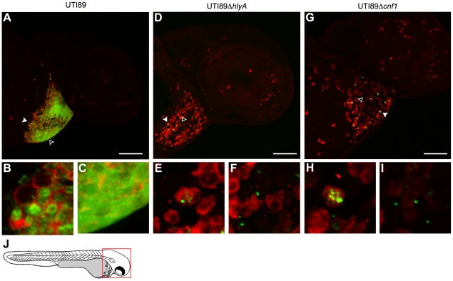
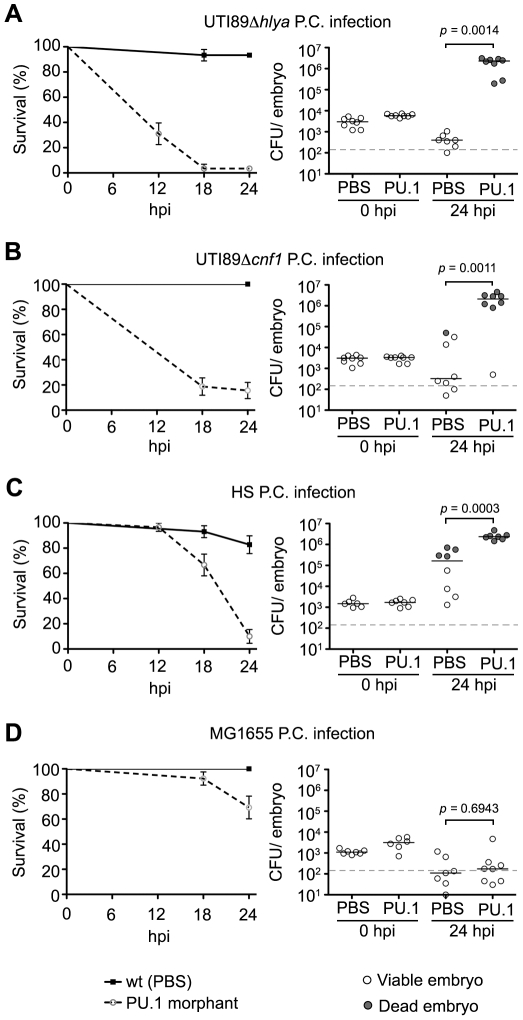
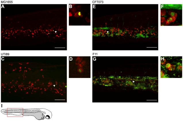
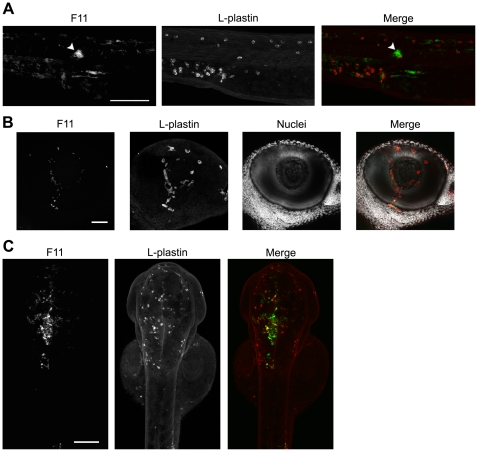
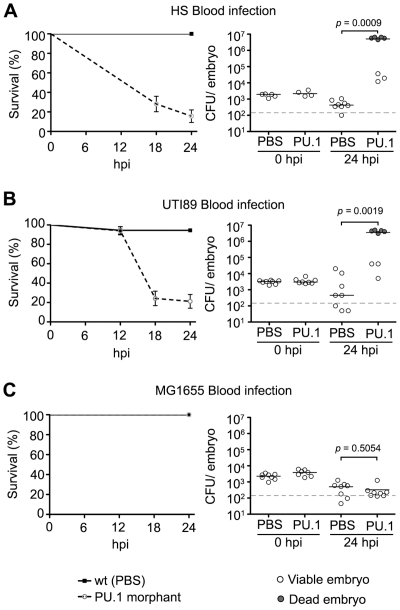
Extraintestinal pathogenic E. coli (ExPEC) cause an array of diseases, including sepsis, neonatal meningitis, and urinary tract infections. Many putative virulence factors that might modulate ExPEC pathogenesis have been identified through sequencing efforts, epidemiology, and gene expression profiling, but few of these genes have been assigned clearly defined functional roles during infection. Using zebrafish embryos as surrogate hosts, we have developed a model system with the ability to resolve diverse virulence phenotypes and niche-specific restrictions among closely related ExPEC isolates during either localized or systemic infections. In side-by-side comparisons of prototypic ExPEC isolates, we observed an unexpectedly high degree of phenotypic diversity that is not readily apparent using more traditional animal hosts. In particular, the capacity of different ExPEC isolates to persist and multiply within the zebrafish host and cause disease was shown to be variably dependent upon two secreted toxins, alpha-hemolysin and cytotoxic necrotizing factor. Both of these toxins appear to function primarily in the neutralization of phagocytes, which are recruited in high numbers to sites of infection where they act as an essential host defense against ExPEC as well as less virulent E. coli strains. These results establish zebrafish as a valuable tool for the elucidation and functional analysis of both ExPEC virulence factors and host defense mechanisms.


Conflict of interest statement

The authors have declared that no competing interests exist.

Figures

Figure 1
Figure 1. Zebrafish survival following infection with ExPEC and related strains.
(A) The pericardial cavities (P.C., top row) or blood (bottom) of 48 hpf embryos were inoculated with PBS containing UTI89, CFT073, ABU 83972, or HS at low (2,000 to 3,500 CFU), medium (4,000 to 6,500 CFU), or high (6,600 to 11,500 CFU) doses. Fish were scored for death at 0, 6, 12, 18, and 24 h post-inoculation (hpi). Data are presented as Kaplan-Meier survival plots with standard error calculated by the Greenwood method. Absence of a tick mark indicates that no deaths were observed at that given time point. (B) Bars show percentage (±standard error) of fish that were killed by ∼5,000 CFU of each strain by 24 h post-inoculation of either the P.C. or blood. n = 30−60 embryos. (C) Bacteria were enumerated from embryos at the indicated times post-inoculation of the P.C. (top) or blood (bottom). Each circle represents bacterial titers from individual embryos that were scored as live (open circles) or dead (shaded circles) prior to homogenization. The dashed line indicates the LOQ.
Figure 2
Figure 2. eBURST diagram of E. coli.
The eBURST algorithm was used to generate a clustering diagram from 2,208 strains from the E. coli MLST database that is maintained by University College Cork. Circles represent single allele profiles (strain types, ST), based on seven genes (adk, fumC, gyrB, icd, mdh, purA, and recA). Size of each circle is proportional to the number of isolates that match a given allele profile. Blue circles are predicted founder genotypes, while red circles represent sub-founder genotypes. Solid lines radiating from founders and sub-founders denote single locus variants. Positions of the 10 sequenced E. coli isolates used in this study are indicated. Bootstrap confidence values for the founder groups associated with the highlighted strains are each 100%, except for the groups containing F11 and HS, which had values of 99% and 93%, respectively.
Figure 3
Figure 3. Differential effects of ExPEC-encoded toxins on zebrafish survival.
(A) and (B), left panels - Zebrafish viability was determined at 0, 6, 12, 18, and 24 hpi of the P.C. with medium doses (4,000–6,500 CFU) of wild type (wt) UTI89, UTI89ΔhlyA, or UTI89ΔhlyA/pSF4000 (+Hly), UTI89Δcnf1, or UTI89Δcnf1/pHLK102 (+CNF1). Right panels - Bacterial burden was determined for UTI89ΔhlyA and UTI89Δcnf1 at the indicated hpi. Embryos were scored as live (open circles) or dead (shaded circles) prior to homogenization. The dashed line denotes the LOQ. (C) Zebrafish survival following inoculation of the P.C. (left) or blood (right) with wt CFT073 or CFT073ΔhlyA. Inoculum sizes used were 2,000–3,500 CFU for P.C. injections and 4,000–6,500 CFU for blood injections. (D) Zebrafish survival after P.C. inoculation with 4,000–6,500 CFU of wt HS (Hly- and CNF1-negative), HS/pSF4000 (+Hly), or HS/pHLK102 (+CNF1). Survival plots (A–D) are presented as Kaplan-Meier survival plots with standard error calculated by the Greenwood method. n = 30−60 embryos for all results.
Figure 4
Figure 4. Phagocyte recruitment and abatement within the zebrafish pericardial cavity.
(A and B) Zebrafish embryos were inoculated via the P.C. with 4,000–6,500 CFU of (A) the K12 strain MG1655 or (B) the ExPEC isolate F11, both carrying pGEN-GFP(LVA) for constitutive expression of destabilized GFP protein (green). Samples were fixed at 0, 6, and 24 hpi and processed for fluorescent confocal microscopy. Phagocytes (red) were detected using L-plastin-specific antibody. For clarity, the merged image at each time point is accompanied by images showing only corresponding single channel signals from bacteria or L-plastin. Each embryo was visualized by stitching together two z-projections generated from 40 to 50 5-µm-thick optical sections. Scale bar = 200 µm. (C) Tg(mpo::GFP) zebrafish embryos, in which GFP expression is under control of the neutrophil-specific MPO promoter, were injected via the P.C. with ∼5,000 CFU of the ExPEC isolate F11. At 4 hpi, samples were fixed and stained using anti-L-plastin antibody to label total phagocytes (red) relative to the GFP-positive neutrophils (green plus red). Bacteria present within the P.C. are not shown. Scale bar = 100 µm. (D) Diagram of a 48 hpf embryo with the area imaged in (C) highlighted by a red box. Embryos shown here are representative. All fish were viable at the time of sacrifice, except the 24 hpi F11-infected fish, which died prior to collection.
Figure 5
Figure 5. Phagocyte localization and bacterial internalization within the pericardial cavity.
Zebrafish embryos were injected via the P.C. with 4,000–6,500 CFU of (A–C) wt UTI89, (D–F) UTI89ΔhlyA, or (G–I) UTI89Δcnf1, each carrying pGEN-GFP(LVA) for constitutive expression of destabilized GFP protein (green). At 6 hpi, samples were fixed and processed for fluorescent confocal microscopy, using anti-L-plastin antibody to label phagocytes (red). Examples of internalized (solid arrowheads) and free extracellular (hollow arrowheads) bacteria in (A, D, and G) are shown at higher magnification in (B, E, and H) and (C, F, and I), respectively. Representative images are shown. All fish were viable at the time of sacrifice. Scale bars = 100 µm. (J) Diagram of a 48 hpf embryo with the area imaged in (A), (D), and (G) highlighted by a red box.
Figure 6
Figure 6. Zebrafish phagocytes are required for resolution of normally non-lethal pericardial E. coli infections.
PU.1 morphants or normal PBS-injected control embryos were inoculated via the P.C. with 4,000–6,500 CFU of (A) UTI89ΔhlyA, (B) UTI89Δcnf1, (C), HS or (D) MG1655. Host survival (left) was evaluated at 0, 6, 12, 18, and 24 hpi and is presented as Kaplan-Meier survival plots with standard error calculated by the Greenwood method. n = 30−60 embryos. (right) Scatter plots show levels of bacterial burden at 0 and 24 hpi in individual embryos that were scored as live (open circles) or dead (shaded circles) at time of collection. The horizontal bars represent median values for each group and the dashed line indicates the LOQ. The P values were determined by Mann-Whitney two-tailed analysis.
Figure 7
Figure 7. Differential growth and phagocytosis of E. coli isolates within the blood.
Zebrafish embryos were infected via the blood with 4,000–6,500 CFU of (A and B) MG1655, (C and D) UTI89, (E and F) CFT073, or (G and H) F11. All bacterial strains carry pGEN-GFP(LVA) for constitutive expression of destabilized GFP (green). At 12 hpi, samples were fixed and phagocytes (red) were labeled using L-plastin-specific antibody for visualization by fluorescent confocal microscopy. Regions highlighted by arrowheads in (A), (C), (E), and (G) are shown further magnified in panels (B), (D), (F), and (H), respectively. All images shown are representative of the pool of embryos imaged. MG1655- and UTI89-infected embryos were viable and healthy in appearance prior to sacrifice for microscopy, whereas fish inoculated with CFT073 or F11 were notably sick and near death at time of collection. Scale bars = 100 µm. (I) Diagram of a 48 hpf embryo with the region imaged in (A), (C), (E), and (G) denoted by a red box.
Figure 8
Figure 8. ExPEC isolates capable of blood colonization can disseminate and persist in a variety of host microenvironments.
F11, carrying pGEN-GFP(LVA) for constitutive expression of destabilized GFP (green), was inoculated into the blood using a dose of 4,000–6,500 CFU. Samples were fixed at 6 or 12 hpi and phagocytes (red) were labeled using L-plastin-specific antibody for visualization by fluorescent confocal microscopy. (A) 20X z-projection of the tail region from an F11-infected embryo at 12 hpi. The arrowhead indicates a commonly observed bacterial microcluster. (B) 40X z-projection of the eye from an embryo at 6 hpi with F11. Hoechst nuclear dye (grey) was used to highlight the anatomical structure of the eye. (C) Dorsal to ventral 10X z-projection of the head region of an embryo at 12 hpi with F11. Scale bars = 100 µm for (A) and (C) and 50 µm for (B).
Figure 9
Figure 9. Zebrafish phagocytes are required for resolution of normally non-lethal E. coli infections in the blood.
PU.1 morphants or normal PBS-injected control embryos were inoculated via the circulation valley with 4,000–6,500 CFU of (A) HS, (B) UTI89, or (C) MG1655. Host survival (left) was evaluated at 0, 6, 12, 18, and 24 hpi and is presented as Kaplan-Meier survival plots with standard error calculated by the Greenwood method. n = 30−60 embryos. Bacterial burden within individual embryos at 0 and 24 hpi is presented as scatter plots on the right. Embryos were scored as live (open circles) or dead (shaded circles) prior to collection. The horizontal lines indicate median values for each group while the dashed line indicates the LOQ. P values were calculated using Mann-Whitney two-tailed analysis.

Similar articles

Cited by

References

    1. Kaper JB, Nataro JP, Mobley HL. Pathogenic Escherichia coli. Nat Rev Microbiol. 2004;2:123–140. - PubMed
    1. Foxman B. Epidemiology of urinary tract infections: incidence, morbidity, and economic costs. Dis Mon. 2003;49:53–70. - PubMed
    1. Russo TA, Johnson JR. Proposal for a new inclusive designation for extraintestinal pathogenic isolates of Escherichia coli: ExPEC. J Infect Dis. 2000;181:1753–1754. - PubMed
    1. Smith JL, Fratamico PM, Gunther NW. Extraintestinal pathogenic Escherichia coli. Foodborne Pathog Dis. 2007;4:134–163. - PubMed
    1. Johnson TJ, Kariyawasam S, Wannemuehler Y, Mangiamele P, Johnson SJ, et al. The genome sequence of avian pathogenic Escherichia coli strain O1:K1:H7 shares strong similarities with human extraintestinal pathogenic E. coli genomes. J Bacteriol. 2007;189:3228–3236. - PMC - PubMed

Publication types