

Nuclear magnetic resonance detects phosphoinositide 3-kinase/Akt-independent traits common to pluripotent murine embryonic stem cells and their malignant counterparts
- PMID: 20019838
- PMCID: PMC2794511
- DOI: 10.1593/neo.09850
Nuclear magnetic resonance detects phosphoinositide 3-kinase/Akt-independent traits common to pluripotent murine embryonic stem cells and their malignant counterparts
Abstract
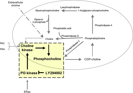
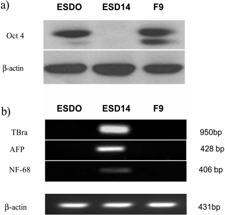
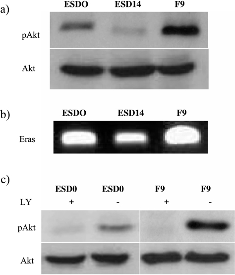
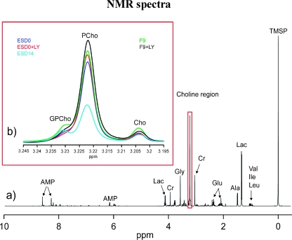
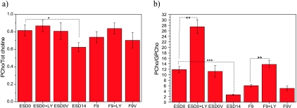
Pluripotent embryonic stem (ES) cells, a potential source of somatic precursors for cell therapies, cause tumors after transplantation. Studies of mammalian carcinogenesis using nuclear magnetic resonance (NMR) spectroscopy have revealed changes in the choline region, particularly increased phosphocholine (PCho) content. High PCho levels in murine ES (mES) cells have recently been attributed to cell pluripotency. The phosphoinositide 3-kinase (PI3K)/Akt pathway has been implicated in tumor-like properties of mES cells. This study aimed to examine a potential link between the metabolic profile associated with choline metabolism of pluripotent mES cells and PI3K/Akt signaling. We used mES (ES-D3) and murine embryonal carcinoma cells (EC-F9) and compared the metabolic profiles of 1) pluripotent mES (ESD0), 2) differentiated mES (ESD14), and 3) pluripotent F9 cells. Involvement of the PI3K/Akt pathway was assessed using LY294002, a selective PI3K inhibitor. Metabolic profiles were characterized in the extracted polar fraction by (1)H NMR spectroscopy. Similarities were found between the levels of choline phospholipid metabolites (PCho/total choline and PCho/glycerophosphocholine [GPCho]) in ESD0 and F9 cell spectra and a greater-than five-fold decrease of the PCho/GPCho ratio associated with mES cell differentiation. LY294002 caused no significant change in relative PCho levels but led to a greater-than two-fold increase in PCho/GPCho ratios. These results suggest that the PCho/GPCho ratio is a metabolic trait shared by pluripotent and malignant cells and that PI3K does not underlie its development. It is likely that the signature identified here in a mouse model may be relevant for safe therapeutic applications of human ES cells.
Figures
Similar articles
-
Metabolic consequences of treatment with AKT inhibitor perifosine in breast cancer cells.NMR Biomed. 2012 Feb;25(2):379-88. doi: 10.1002/nbm.1764. Epub 2011 Aug 23. NMR Biomed. 2012. PMID: 22253088 Free PMC article.
-
Reduced phosphocholine and hyperpolarized lactate provide magnetic resonance biomarkers of PI3K/Akt/mTOR inhibition in glioblastoma.Neuro Oncol. 2012 Mar;14(3):315-25. doi: 10.1093/neuonc/nor209. Epub 2011 Dec 12. Neuro Oncol. 2012. PMID: 22156546 Free PMC article.
-
Akt suppresses DLK for maintaining self-renewal of mouse embryonic stem cells.Cell Cycle. 2015;14(8):1207-17. doi: 10.1080/15384101.2015.1014144. Cell Cycle. 2015. PMID: 25802931 Free PMC article.
-
[Progress on PI3K/Akt signaling pathway regulating self-renewal and pluripotency of embryonic stem cells].Sheng Li Xue Bao. 2014 Apr 25;66(2):223-30. Sheng Li Xue Bao. 2014. PMID: 24777414 Review. Chinese.
-
Small-molecule inhibitors of the PI3K signaling network.Future Med Chem. 2011 Apr;3(5):549-65. doi: 10.4155/fmc.11.12. Future Med Chem. 2011. PMID: 21526896 Free PMC article. Review.
Cited by
-
Choline kinase inhibition in rheumatoid arthritis.Ann Rheum Dis. 2015 Jul;74(7):1399-407. doi: 10.1136/annrheumdis-2014-205696. Epub 2014 Oct 1. Ann Rheum Dis. 2015. PMID: 25274633 Free PMC article.
-
Metabolomic high-content nuclear magnetic resonance-based drug screening of a kinase inhibitor library.Nat Commun. 2011 Nov 22;2:545. doi: 10.1038/ncomms1562. Nat Commun. 2011. PMID: 22109519 Free PMC article.
-
The interconnectedness of cancer cell signaling.Neoplasia. 2011 Dec;13(12):1183-93. doi: 10.1593/neo.111746. Neoplasia. 2011. PMID: 22241964 Free PMC article.
-
Large-scale transcriptome profiles reveal robust 20-signatures metabolic prediction models and novel role of G6PC in clear cell renal cell carcinoma.J Cell Mol Med. 2020 Aug;24(16):9012-9027. doi: 10.1111/jcmm.15536. Epub 2020 Jun 21. J Cell Mol Med. 2020. PMID: 32567187 Free PMC article.
-
Hypoxia triggers major metabolic changes in AML cells without altering indomethacin-induced TCA cycle deregulation.ACS Chem Biol. 2011 Feb 18;6(2):169-75. doi: 10.1021/cb900300j. Epub 2010 Oct 18. ACS Chem Biol. 2011. PMID: 20886892 Free PMC article.
References
-
- Takahashi K, Ichisaka T, Yamanaka S. Identification of genes involved in tumor-like properties of embryonic stem cells. Methods Mol Biol. 2006;329:449–458. - PubMed
-
- Wang Y, Armstrong SA. Cancer: inappropriate expression of stem cell programs? Cell Stem Cell. 2008;2:297–299. - PubMed
-
- Takahashi K, Mitsui K, Yamanaka S. Role of ERas in promoting tumour-like properties in mouse embryonic stem cells. Nature. 2003;423:541–545. - PubMed
-
- Turner WS, Seagle C, Galanko JA, Favorov O, Prestwich GD, Macdonald JM, Reid LM. Nuclear magnetic resonance metabolomic footprinting of human hepatic stem cells and hepatoblasts cultured in hyaluronan-matrix hydrogels. Stem Cells. 2008;26:1547–1555. - PubMed
Publication types
MeSH terms
Substances
Grants and funding
LinkOut - more resources
Full Text Sources
Miscellaneous