

Chemotherapeutic drug-induced ABCG2 promoter demethylation as a novel mechanism of acquired multidrug resistance
- PMID: 20019844
- PMCID: PMC2794517
- DOI: 10.1593/neo.91314
Chemotherapeutic drug-induced ABCG2 promoter demethylation as a novel mechanism of acquired multidrug resistance
Abstract
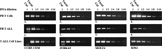
ABCG2 is an efflux transporter conferring multidrug resistance (MDR) on cancer cells. However, the initial molecular events leading to its up-regulation in MDR tumor cells are poorly understood. Herein, we explored the impact of drug treatment on the methylation status of the ABCG2 promoter and consequent reactivation of ABCG2 gene expression in parental tumor cell lines and their MDR sublines. We demonstrate that ABCG2 promoter methylation is common in T-cell acute lymphoblastic leukemia (T-ALL) lines, also present in primary T-ALL lymphoblast specimens. Furthermore, drug selection with sulfasalazine and topotecan induced a complete demethylation of the ABCG2 promoter in the T-ALL and ovarian carcinoma model cell lines CCRF-CEM and IGROV1, respectively. This resulted in a dramatic induction of ABCG2 messenger RNA levels (235- and 743-fold, respectively) and consequent acquisition of an ABCG2-dependent MDR phenotype. Quantitative genomic polymerase chain reaction and ABCG2 promoter-luciferase reporter assay did not reveal ABCG2 gene amplification or differential transcriptional trans-activation, which could account for ABCG2 up-regulation in these MDR cells. Remarkably, mimicking cytotoxic bolus drug treatment through 12- to 24-hour pulse exposure of ABCG2-silenced leukemia cells, to clinically relevant concentrations of the chemotherapeutic agents daunorubicin and mitoxantrone, resulted in a marked transcriptional up-regulation of ABCG2. Our findings establish that antitumor drug-induced epigenetic reactivation of ABCG2 gene expression in cancer cells is an early molecular event leading to MDR. These findings have important implications for the emergence, clonal selection, and expansion of malignant cells with the MDR phenotype during chemotherapy.
Figures


Similar articles
-
ABCG2 expression, function, and promoter methylation in human multiple myeloma.Blood. 2006 Dec 1;108(12):3881-9. doi: 10.1182/blood-2005-10-009084. Epub 2006 Aug 17. Blood. 2006. PMID: 16917002 Free PMC article. Clinical Trial.
-
Single-step doxorubicin-selected cancer cells overexpress the ABCG2 drug transporter through epigenetic changes.Br J Cancer. 2008 May 6;98(9):1515-24. doi: 10.1038/sj.bjc.6604334. Epub 2008 Apr 1. Br J Cancer. 2008. PMID: 18382425 Free PMC article.
-
Expression of ABCG2 (BCRP) is regulated by Nrf2 in cancer cells that confers side population and chemoresistance phenotype.Mol Cancer Ther. 2010 Aug;9(8):2365-76. doi: 10.1158/1535-7163.MCT-10-0108. Epub 2010 Aug 3. Mol Cancer Ther. 2010. PMID: 20682644 Free PMC article.
-
Multidrug resistance in cancer chemotherapy and xenobiotic protection mediated by the half ATP-binding cassette transporter ABCG2.Curr Med Chem Anticancer Agents. 2004 Jan;4(1):31-42. doi: 10.2174/1568011043482205. Curr Med Chem Anticancer Agents. 2004. PMID: 14754410 Review.
-
ABCG2 in Acute Myeloid Leukemia: Old and New Perspectives.Int J Mol Sci. 2023 Apr 12;24(8):7147. doi: 10.3390/ijms24087147. Int J Mol Sci. 2023. PMID: 37108308 Free PMC article. Review.
Cited by
-
Integration and bioinformatics analysis of DNA-methylated genes associated with drug resistance in ovarian cancer.Oncol Lett. 2016 Jul;12(1):157-166. doi: 10.3892/ol.2016.4608. Epub 2016 May 18. Oncol Lett. 2016. PMID: 27347118 Free PMC article.
-
An 8‑gene signature predicts the prognosis of cervical cancer following radiotherapy.Mol Med Rep. 2019 Oct;20(4):2990-3002. doi: 10.3892/mmr.2019.10535. Epub 2019 Jul 29. Mol Med Rep. 2019. PMID: 31432147 Free PMC article.
-
Development of precision medicine approaches based on inter-individual variability of BCRP/ABCG2.Acta Pharm Sin B. 2019 Jul;9(4):659-674. doi: 10.1016/j.apsb.2019.01.007. Epub 2019 Jan 15. Acta Pharm Sin B. 2019. PMID: 31384528 Free PMC article. Review.
-
Targeting ABCG2 transporter to enhance 5-aminolevulinic acid for tumor visualization and photodynamic therapy.Biochem Pharmacol. 2023 Nov;217:115851. doi: 10.1016/j.bcp.2023.115851. Epub 2023 Oct 17. Biochem Pharmacol. 2023. PMID: 37858868 Free PMC article. Review.
-
Abnormal methylation characteristics predict chemoresistance and poor prognosis in advanced high-grade serous ovarian cancer.Clin Epigenetics. 2021 Jul 21;13(1):141. doi: 10.1186/s13148-021-01133-2. Clin Epigenetics. 2021. PMID: 34289901 Free PMC article.
References
-
- Szakacs G, Paterson JK, Ludwig JA, Booth-Genthe C, Gottesman MM. Targeting multidrug resistance in cancer. Nat Rev Drug Discov. 2006;5:219–234. - PubMed
-
- Borst P, Elferink RO. Mammalian ABC transporters in health and disease. Annu Rev Biochem. 2002;71:537–592. - PubMed
-
- Dean M, Rzhetsky A, Allikmets R. The human ATP-binding cassette (ABC) transporter superfamily. Genome Res. 2001;11:1156–1166. - PubMed
-
- Borst P, Evers R, Kool M, Wijnholds J. A family of drug transporters: the multidrug resistance-associated proteins. J Natl Cancer Inst. 2000;92:1295–1302. - PubMed
-
- Deeley RG, Westlake C, Cole SP. Transmembrane transport of endo- and xenobiotics by mammalian ATP-binding cassette multidrug resistance proteins. Physiol Rev. 2006;86:849–899. - PubMed
Publication types
MeSH terms
Substances
LinkOut - more resources
Full Text Sources