. 2009 Dec 12:15:2751-61.

Retinal phototoxicity in a novel murine model of intraocular lens implantation

Affiliations

Retinal phototoxicity in a novel murine model of intraocular lens implantation

Toshihide Kurihara et al. Mol Vis. .

Abstract

Purpose: To establish a novel murine intraocular lens (IOL) implantation model to study the protective effects of colored-IOLs against retinal phototoxicity.

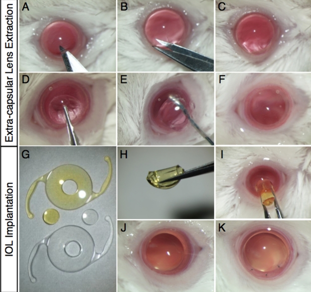
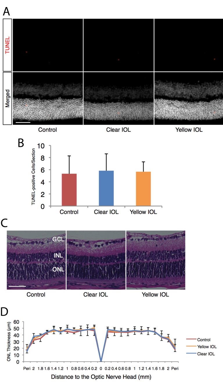
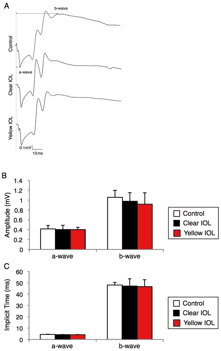
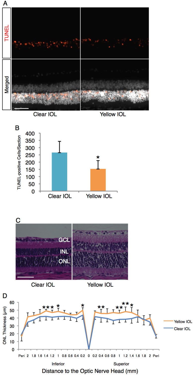
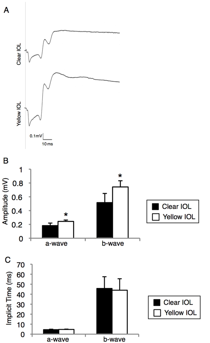
Methods: Two-millimeter diameter IOL buttons were created from IOLs for clinical use. Extra-capsular crystalline lens extraction and IOL implantation were performed in BALB/c mice using a technique similar to human cataract surgery. For light exposure experiments, mice were exposed to 5,000 LUX of white light for 24 h on the day after surgery. To investigate the protective effects of yellow IOL against light exposure, ERG measurements were conducted in vivo, followed by TdT-mediated dUTP Nick-End Labeling (TUNEL) and outer nuclear layer (ONL) thickness measurement of retinal tissue in yellow or clear IOL-implanted mice and control mice without surgery.

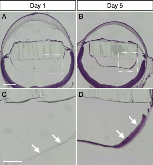
Results: IOLs were successfully implanted in all animals, and IOL buttons without haptics were well stabilized in the capsular bag. Murine eyes developed posterior capsule opacification (PCO) after IOL implantation by postoperative day 5 at the latest. In contrast to the clear IOL-implanted animals stimulated by light exposure, the yellow IOL-implanted animals had significantly reduced numbers of TUNEL-positive cells and retained thickness of the ONL. The ERG showed that yellow IOL implantation prevents a decrease of amplitude in both the a-wave and b-wave compared with clear IOL implantation.

Conclusions: We established a new animal model of IOL implantation and demonstrated the protective effects of colored-IOL against retinal phototoxicity after cataract surgery.


Figures

Figure 1
Figure 1
Extra-capsular crystalline lens extraction and IOL implantation in murine eyes. A-C: Corneal incision with a surgical knife and Vannas scissors. D: Continuous curvilinear capsulorrhexis with forceps. E: Lens extraction with hydrodissection. F: Inflation of the capsular bag with a viscoelastic substance. G: Fabricated IOL buttons (2 mm diameter). H, I: IOL insertion. J, K: Removal of the viscoelastic substance and closure of the corneal wound with interrupted 11–0 nylon sutures.
Figure 2
Figure 2
Development of posterior capsular opacity in murine IOL-implanted eyes. Representative micrographs of murine IOL-implanted eyes, stained with hematoxylin and eosin on post-operative day 1 (A) or day 5 (B) and high-magnification images of the posterior lens capsule (C, D; square area marked in A and B, respectively). The arrows depict lens epithelial cells migrating underneath the posterior capsule on post-operative day 5 (D), whereas no cells were seen on post-operative day 1 (C). The bar shown in A represents 500 µm and applies for (B) as well. The bar shown in C represents 200 µm and applies for (D) as well.
Figure 3
Figure 3
Morphological assessment of sensory retina in murine IOL-implanted eyes and control eyes without surgery. Apoptotic photoreceptors and ONL thickness after IOL implantation were assessed under dim light conditions (5 LUX, 12 h on/off). A: (Upper) Representative images of TUNEL staining for retinal sections 1 mm superior to the optic nerve head at postoperative day 3. (Lower) Merged images. Nuclei were counterstained with Hoechst 33258. Bar=50 µm. B: Quantification of TUNEL-positive cells in the ONL of each section, including the optic nerve head. Values are mean ±SD (n=6 in each group). C: Representative images of hematoxylin and eosin staining for retinal sections 1 mm superior to the optic nerve head at postoperative day 7. Bar represents 50 µm. D: ONL thickness of control or IOL-implanted eyes. Values are mean ±SD (n=5 to 6 in each group).
Figure 4
Figure 4
Functional assessment of sensory retina in murine IOL-implanted eyes. ERG was measured under dim (5 LUX, 12 h on/off) light at 7 days after IOL implantation. A: Representative wave responses from control or IOL-implanted eyes. B, C: Quantification of amplitude (B) or implicit time (C) of a-wave and b-wave. Values are mean ±SD (n=5 in each group).
Figure 5
Figure 5
Impact of yellow IOL on the light-induced retinal morphological changes. A: (Upper) Representative images of TUNEL staining for retinal sections 1 mm superior to the optic nerve head on postoperative day 3 (2 days after the light exposure [5,000 LUX, 24 h]). (Lower) Merged images. Nuclei were counterstained with Hoechst 33258. Bar represents 50 µm. B: Quantification of TUNEL-positive cells in the ONL of each section, including the optic nerve head. Values are mean ±SD (n=6 in each group; *p<0.05). C: Representative images of hematoxylin and eosin staining for retinal sections 1 mm superior to the optic nerve head on postoperative day 7 (6 days after the light exposure [5,000 LUX, 24 h]). Bar represents 50 µm. D: ONL thickness of IOL-implanted eyes after light exposure. Values are mean ±SD (n=5 to 7; *p<0.05, **p<0.01).
Figure 6
Figure 6
Protective effects of yellow IOL against light-induced retinal functional damage. ERG was measured at postoperative day 7 (6 days after the light exposure [5,000 LUX, 24 h]) in IOL implanted-mice. A: Representative wave responses from clear or yellow IOL-implanted eyes. B, C: Quantification of amplitude (B) or implicit time (C) of a-waves and b-waves from each group (n=5 in each group, respectively; *p<0.05).

Similar articles

Cited by

References

    1. Clark DS. Posterior capsule opacification. Curr Opin Ophthalmol. 2000;11:56–64. - PubMed
    1. Ray S, D'Amico DJ. Pseudophakic cystoid macular edema. Semin Ophthalmol. 2002;17:167–80. - PubMed
    1. Bockelbrink A, Roll S, Ruether K, Rasch A, Greiner W, Willich SN. Cataract surgery and the development or progression of age-related macular degeneration: a systematic review. Surv Ophthalmol. 2008;53:359–67. - PubMed
    1. Congdon N, Vingerling JR, Klein BE, West S, Friedman DS, Kempen J, O'Colmain B, Wu SY, Taylor HR, Eye Diseases Prevalence Research Group. Prevalence of cataract and pseudophakia/aphakia among adults in the United States. Arch Ophthalmol. 2004;122:487–94. - PubMed
    1. Wenzel A, Grimm C, Samardzija M, Reme CE. Molecular mechanisms of light-induced photoreceptor apoptosis and neuroprotection for retinal degeneration. Prog Retin Eye Res. 2005;24:275–306. - PubMed

Publication types

LinkOut - more resources