

FcRn receptor-mediated pharmacokinetics of therapeutic IgG in the eye
- PMID: 20019892
- PMCID: PMC2794657
FcRn receptor-mediated pharmacokinetics of therapeutic IgG in the eye
Abstract
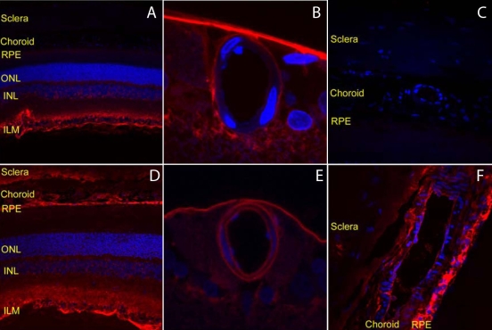
Purpose: The goal of this study was to determine the role of the neonatal Fc (FcRn) receptor in eliminating intravitreally administered full-length immunoglobulin G (IgG) across the blood-retinal barrier.
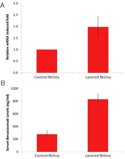
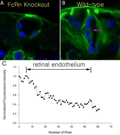
Methods: FcRn receptor expression in normal and laser photocoagulated retinas was compared quantitatively by real-time RT-PCR. The distribution of intravitreally administered full-length IgG was investigated and compared in wild-type and FcRn knockout mouse eyes as well as normal and laser-photocoagulated rat eyes at several time points. Additionally, the pharmacokinetics of intravitreally injected full-length IgG and chicken immunoglobulin Y (IgY) was compared in the normal rat retina.
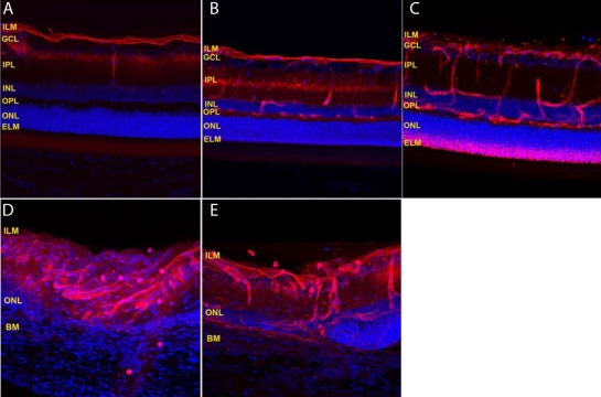
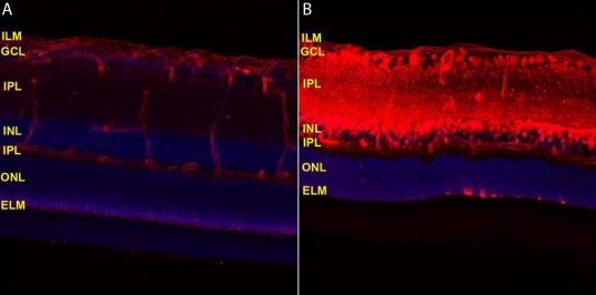
Results: Intravitreally administered full-length IgG overcame the inner limiting membrane and diffused into the deeper retinal structures in both normal and laser-photocoagulated retinas. Interestingly, IgG was eliminated across the blood-retinal barrier into the blood system in the normal retina, whereas IgY was not. In addition, full-length IgGs did not penetrate across the blood-retinal barrier in the FcRn knockout mouse. Intravitreally injected IgGs were eliminated into the blood system more rapidly in laser-photocoagulated eyes when compared to normal control eyes because of FcRn receptor upregulation in the laser-photocoagulated retina.
Conclusions: FcRn plays an important role in eliminating intravitreally administered full-length IgGs across the blood-retina barrier into the systemic blood system.
Figures
Similar articles
-
Expression of neonatal Fc receptor in the eye.Invest Ophthalmol Vis Sci. 2014 Mar 19;55(3):1607-15. doi: 10.1167/iovs.13-12574. Invest Ophthalmol Vis Sci. 2014. PMID: 24550358 Free PMC article.
-
The role of Fc-receptors in the uptake and transport of therapeutic antibodies in the retinal pigment epithelium.Exp Eye Res. 2016 Apr;145:187-205. doi: 10.1016/j.exer.2015.12.013. Epub 2016 Jan 8. Exp Eye Res. 2016. PMID: 26773870
-
Mapping of the neonatal Fc receptor in the rodent eye.Invest Ophthalmol Vis Sci. 2008 May;49(5):2025-9. doi: 10.1167/iovs.07-0871. Invest Ophthalmol Vis Sci. 2008. PMID: 18436836 Free PMC article.
-
Neonatal Fc receptor (FcRn): a novel target for therapeutic antibodies and antibody engineering.J Drug Target. 2014 May;22(4):269-78. doi: 10.3109/1061186X.2013.875030. Epub 2014 Jan 9. J Drug Target. 2014. PMID: 24404896 Review.
-
Neonatal Fc receptor in human immunity: Function and role in therapeutic intervention.J Allergy Clin Immunol. 2020 Sep;146(3):467-478. doi: 10.1016/j.jaci.2020.07.015. J Allergy Clin Immunol. 2020. PMID: 32896307 Review.
Cited by
-
Targeting key angiogenic pathways with a bispecific CrossMAb optimized for neovascular eye diseases.EMBO Mol Med. 2016 Nov 2;8(11):1265-1288. doi: 10.15252/emmm.201505889. Print 2016 Nov. EMBO Mol Med. 2016. PMID: 27742718 Free PMC article.
-
In Translation: FcRn across the Therapeutic Spectrum.Int J Mol Sci. 2021 Mar 17;22(6):3048. doi: 10.3390/ijms22063048. Int J Mol Sci. 2021. PMID: 33802650 Free PMC article. Review.
-
Neonatal Fc receptor: from immunity to therapeutics.J Clin Immunol. 2010 Nov;30(6):777-89. doi: 10.1007/s10875-010-9468-4. Epub 2010 Oct 1. J Clin Immunol. 2010. PMID: 20886282 Free PMC article. Review.
-
Anti-VEGF-refractory exudative age-related macular degeneration: differential response according to features on optical coherence tomography.Korean J Ophthalmol. 2013 Dec;27(6):425-32. doi: 10.3341/kjo.2013.27.6.425. Epub 2013 Nov 15. Korean J Ophthalmol. 2013. PMID: 24311928 Free PMC article.
-
Clinical use of Bevacizumab in treating refractory glaucoma.J Med Life. 2015 Jan-Mar;8(1):8-12. J Med Life. 2015. PMID: 25914729 Free PMC article. Review.
References
-
- Lobo ED, Hansen RJ, Balthasar JP. Antibody pharmacokinetics and pharmacodynamics. J Pharm Sci. 2004;93:2645–68. - PubMed
-
- Heiduschka P, Fietz H, Hofmeister S, Schultheiss S, Mack AF, Peters S, Ziemssen F, Niggemann B, Julien S, Bartz-Schmidt KU, Schraermeyer U, Tübingen Bevacizumab Study Group. Penetration of bevacizumab through the retina after intravitreal injection in the monkey. Invest Ophthalmol Vis Sci. 2007;48:2814–23. - PubMed
-
- Hosoya K, Tomi M. Advances in the cell biology of transport via the inner blood-retinal barrier: establishment of cell lines and transport functions. Biol Pharm Bull. 2005;28:1–8. - PubMed
-
- Stewart PA, Tuor UI. Blood-eye barriers in the rat: correlation of ultrastructure with function. J Comp Neurol. 1994;340:566–76. - PubMed
Publication types
MeSH terms
Substances
LinkOut - more resources
Full Text Sources
Other Literature Sources
Medical