. 2010 Feb 15;518(4):493-512.
doi: 10.1002/cne.22233.

Distribution of androgen receptor mRNA expression in vocal, auditory, and neuroendocrine circuits in a teleost fish

Affiliations

Distribution of androgen receptor mRNA expression in vocal, auditory, and neuroendocrine circuits in a teleost fish

Paul M Forlano et al. J Comp Neurol. .

Abstract

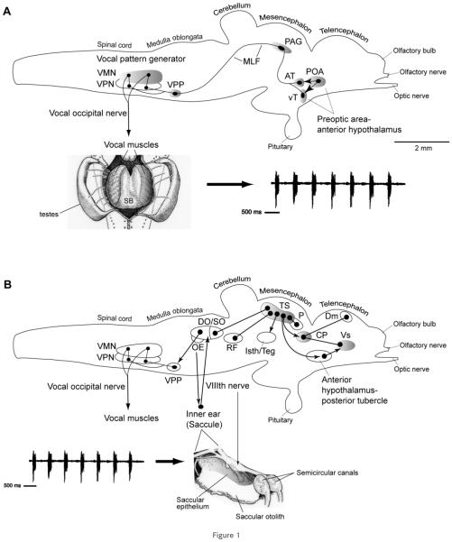
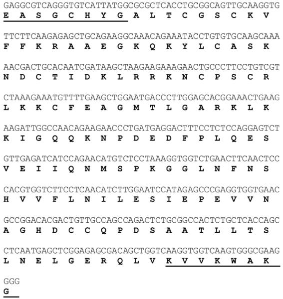
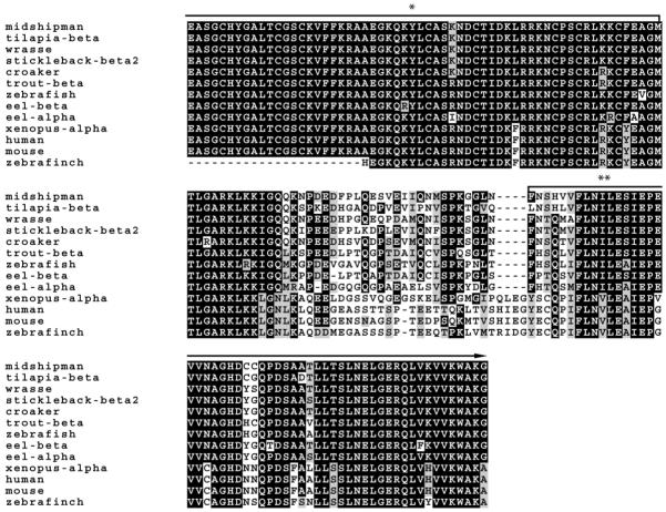
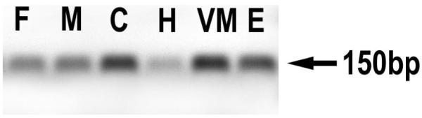
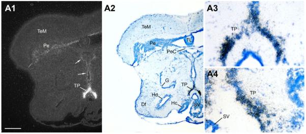
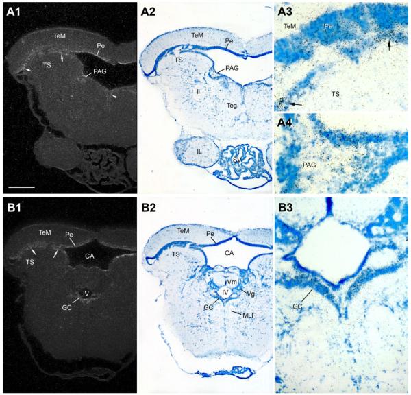
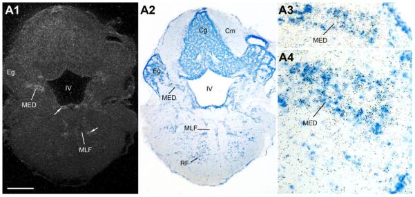
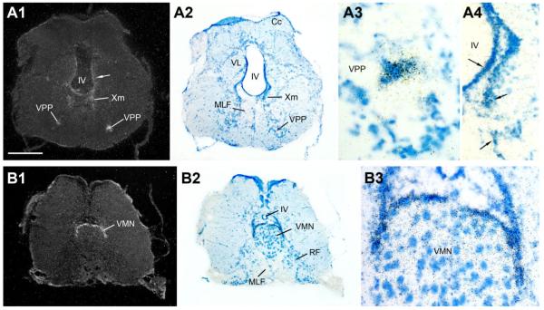
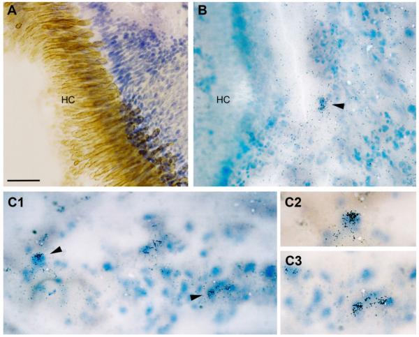
Across all major vertebrate groups, androgen receptors (ARs) have been identified in neural circuits that shape reproductive-related behaviors, including vocalization. The vocal control network of teleost fishes presents an archetypal example of how a vertebrate nervous system produces social, context-dependent sounds. We cloned a partial cDNA of AR that was used to generate specific probes to localize AR expression throughout the central nervous system of the vocal plainfin midshipman fish (Porichthys notatus). In the forebrain, AR mRNA is abundant in proposed homologs of the mammalian striatum and amygdala, and in anterior and posterior parvocellular and magnocellular nuclei of the preoptic area, nucleus preglomerulosus, and posterior, ventral and anterior tuberal nuclei of the hypothalamus. Many of these nuclei are part of the known vocal and auditory circuitry in midshipman. The midbrain periaqueductal gray, an essential link between forebrain and hindbrain vocal circuitry, and the lateral line recipient nucleus medialis in the rostral hindbrain also express abundant AR mRNA. In the caudal hindbrain-spinal vocal circuit, high AR mRNA is found in the vocal prepacemaker nucleus and along the dorsal periphery of the vocal motor nucleus congruent with the known pattern of expression of aromatase-containing glial cells. Additionally, abundant AR mRNA expression is shown for the first time in the inner ear of a vertebrate. The distribution of AR mRNA strongly supports the role of androgens as modulators of behaviorally defined vocal, auditory, and neuroendocrine circuits in teleost fish and vertebrates in general.


Figures

Figure 1
Figure 1
Side views of the brain showing vocal motor (A) and central auditory (B) systems in batrachoidid fish (midshipman and toadfish) (modified from Bass and McKibben, 2003; Kittelberger et al., 2006). Solid dots represent somata, and lines represent axonal projection pathways. Two connected dots indicate reciprocal connections. Shaded areas indicate localization of AR expression. A: Descending vocal motor pathways (see Bass and Baker, 1990; Bass et al., 1994; Goodson and Bass, 2002; Kittelberger et al., 2006). Preoptic (POA) and ventral (vT) and anterior (AT) tuberal nuclei in the hypothalamic forebrain project to the periaqueductal gray (PAG) in the midbrain which then connects to the vocal pattern generator (VPG) in the hindbrain-spinal cord. The VPG consists of vocal prepacemaker (VPP), pacemaker (VPN), and motor (VMN) nuclei. The VMN projects directly via occipital nerve roots to sound-producing muscles on the swim bladder (shown in ventral, dissected view). The arrow points to a vocalization produced by the swim bladder, a typical “grunt train” often produced during nest defense by a type I male (Brantley and Bass, 1994). B: Central auditory system (see Bass et al., 2000, 2001). Natural sounds, represented by a “grunt train,” are detected by the inner ear which projects via the VIIIth nerve to octaval nuclei in the hindbrain and further to the auditory midbrain torus semicircularis (TS). Shown are nuclei interconnected with TS. The dorsal thalamic central posterior nucleus (CP) contains reciprocal connections to the dorsomedial (Dm) and ventral supracommissural (Vs) telencephalon which also receives input from the anterior hypothalamus-posterior tubercle (for nomenclature, see Braford and Northcutt, 1983). TS and CP also connect to vocal motor nuclei in the forebrain (anterior hypothalamus and POA) and midbrain (PAG and isthmal/tegmentum), while auditory-recipient octaval nuclei in the hindbrain connect to the VPG (also see Bass et al., 1994; Goodson and Bass, 2002).
Figure 2
Figure 2
Nucleotide and deduced amino acid sequence of AR partial cDNA clone isolated from midshipman brain. Underlined amino acids and basepairs correspond to sequence derived from degenerate primers.
Figure 3
Figure 3
Alignment of deduced amino acid sequences of midshipman with other representative vertebrate ARs: teleost (tilapia, wrasse, stickleback, croaker, trout, zebrafish, and eel), amphibian (Xenopus), mammalian (human and mouse) and avian (zebra finch). Black and gray shading indicate identical and similar amino acids to midshipman AR, respectively. The first and last eight amino acids are conserved regions to which degenerate primers were made for RT-PCR. The entire sequence falls within the DNA (*) and ligand binding domain (**) and is up to 90% conserved with other teleosts.
Figure 4
Figure 4
Using midshipman-specific primers, a predicted 150-bp product containing AR transcripts was identified by RT-PCR in a forebrain region (F), a middle CNS region including midbrain and caudal diencephalon (M), the cerebellum (C), hindbrain and rostral spinal cord (H), vocal muscle (VM), and saccular epithelium of the inner ear (E) of individual adult midshipman. Shown is a representative type I male.
Figure 5
Figure 5
Dorsal view of exposed brain and inner ear of a midshipman indicating levels (rostral-caudal) of transverse sections for the brain atlas of androgen receptor distribution shown in Figures 6-12. C, cerebellum; M, midbrain; OB, olfactory bulb; OC, occipital nerve roots; T, telencephalon; SE, saccular epithelium of the inner ear; VIII, eighth nerve. Modified from Forlano et al. (2005). Scale bar = 1.5 mm.
Figure 6
Figure 6
AR mRNA expression in the ventral telencephalon and rostral preoptic area. In this figure and in Figures 6-12, neuroanatomical localization of in situ hybridization signal is shown for the same section in low-magnification darkfield (left) and brightfield Nissl-stained sections (middle) and higher-magnification brightfield (right). A13: Robust AR expression in dorsal nucleus of area ventralis (Vd). B1B3: High AR mRNA expression in the supracommissural nucleus of area ventralis (Vs) and anterior parvocellular preoptic area (PPa), part of the forebrain vocal-acoustic complex (see Fig. 1A,B; Table 1). C1C4: Strong AR expression in the dorsal pallium in the central zone of area dorsalis (Dc) as well as throughout the extent of the PPa and the postcommissural nucleus of area ventralis (Vp). Scale bar = 500 μm for panels 1, 2 in A–C; 100 μm, 200 μm, and 100 μm panels in A3, B3, and C3–C4, respectively. [Color figure can be viewed in the online issue, which is available at www.interscience.wiley.com.]
Figure 7
Figure 7
AR mRNA expression in the dorsal telencephalon and caudal preoptic area. A1A5: The posterior (p) and central medial (cm) divisions of the medial zone of area dorsalis (Dm-p and Dm-cm), the intermediate nucleus of area ventralis (Vi), and the gigantocellular division of the magnocellular preoptic area (PMg) show strong AR mRNA expression. B1B4: In addition to strong hybridization signal in the dorsal pallium (Dm-p, Dm-cm) and ventricular zone adjacent to PMg, the ventral tuberal hypothalamus (vT), part of the descending vocal motor pathway (see Fig. 1), shows robust AR expression. Scale bar = 500 μm for panels 1, 2 in A,B; 100 μm for A3–A5 and B3–B4. [Color figure can be viewed in the online issue, which is available at www.interscience.wiley.com.]
Figure 8
Figure 8
AR mRNA expression in the diencephalon and rostral hypothalamus. A1A3: Dorsal (Hd) and ventrolateral periventricular (Hv) nuclei of the hypothalamus show abundant AR; lower levels are found in the anterior tuberal nucleus (AT), part of the forebrain vocal-acoustic complex (see Fig. 1). B1B4: Diffuse AR mRNA expression is found in the auditory central posterior nucleus (CP, see Fig. 1), the dorsal posterior (DPo) nucleus of the thalamus, and the periventricular layer of the midbrain tectum (Pe), as well as along the third ventricle (III) including the periventricular posterior tuberal nucleus (TPp, also see A). Scale bar = 500 μm for panels 1, 2 in A,B; 200 μm and 100 μm for A3 and B3–B4, respectively. [Color figure can be viewed in the online issue, which is available at www.interscience.wiley.com.]
Figure 9
Figure 9
A1A4: The posterior tuberal nucleus (TP), shown in high magnification at two levels, has perhaps the highest abundance of AR mRNA in the central nervous system. Also note consistent signal (arrows) along the ventricular midline in the thalamus (midline) and in the periventricular cell layer of the tectum (Pe). Scale bar = 500 μm for panels A1–A2; 100 μm for A3–A4. [Color figure can be viewed in the online issue, which is available at www.interscience.wiley.com.]
Figure 10
Figure 10
AR mRNA expression at two midbrain levels. A1A4: Hybridization signal in the periaqueductal gray (PAG), part of the descending vocal motor pathway (see Fig. 1), and the periventricular torus semicircularis (TS, arrows), tectum (Pe) and dorsal tegmental midline (arrowhead). B1B3: AR mRNA expression in a more caudal section of the midbrain which also demonstrates signal in periventricular TS (arrows), tectum’s Pe, and griseum centrale (GC). Scale bar = 500 μm for panels 1, 2 in A,B; 100 μm and 200 μm for A3–A4 and B3, respectively. [Color figure can be viewed in the online issue, which is available at www.interscience.wiley.com.]
Figure 11
Figure 11
A1A4: AR mRNA expression in the lateral line recipient nucleus medialis (MED). Also notice diffuse signal along the fourth ventricle (IV) and lateral to the medial longitudinal fasciculus (MLF) (arrows). Scale bar = 500 μm for A1–A2; 200 μm and 100 μm for A3 and A4, respectively. [Color figure can be viewed in the online issue, which is available at www.interscience.wiley.com.]
Figure 12
Figure 12
AR mRNA expression in the vocal, hindbrain-spinal cord pattern generator. A1A4: The vocal prepacemaker nucleus (VPP) shows robust AR expression; also consistent but diffuse mRNA signal found along the ventricle (IV). B1B3: Strong AR mRNA expression forms a cap around the dorsal periphery of the vocal motor nucleus (VMN). Scale bar = 500 μm for panels 1, 2 in A,B; 100 μm in A3; 200 μm in A4,B3. [Color figure can be viewed in the online issue, which is available at www.interscience.wiley.com.]
Figure 13
Figure 13
AR mRNA expression in the inner ear of female midshipman. A: The hair cell layer (HC) of the sensory epithelium of the saccule is delineated by a hair cell-specific antibody (brown), overlaying Nissl stained support cells (see Forlano et al., 2005, for methods). B: Arrowhead indicates AR mRNA (clusters of silver grains) over cells just outside the HC layer. C1C3: Examples of hybridization specificity in the inner ear in a similar location as B. C2C3: Higher magnification of cells indicated by arrowheads in C1. Scale bar = 100 μm in A,B; 50 μm in C1; 30 μm in C2–C3. [Color figure can be viewed in the online issue, which is available at www.interscience.wiley.com.]

Similar articles

Cited by

References

    1. Ball GF, Riters LV, Balthazart J. Neuroendocrinology of song behavior and avian brain plasticity: multiple sites of action of sex steroid hormones. Front Neuroendocrinol. 2002;23:137–178. - PubMed
    1. Bass AH. Shaping brain sexuality. Am Sci. 1996;84:352–363.
    1. Bass AH. Steroid-dependent plasticity of vocal motor systems: novel insights from teleost fish. Brain Res Rev. 2008;57:299–308. - PubMed
    1. Bass AH, Baker R. Sexual dimorphisms in the vocal control system of a teleost fish: morphology of physiologically identified neurons. J Neurobiol. 1990;21:1155–1168. - PubMed
    1. Bass AH, Baker R. Phenotypic specification of hindbrain rhombomeres and the origins of rhythmic circuits in vertebrates. Brain Behav Evol. 1997;50:3–16. - PMC - PubMed

Publication types

MeSH terms