

Innate immune evasion strategies of influenza viruses
- PMID: 20020828
- PMCID: PMC2820251
- DOI: 10.2217/fmb.09.108
Innate immune evasion strategies of influenza viruses
Abstract
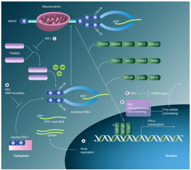
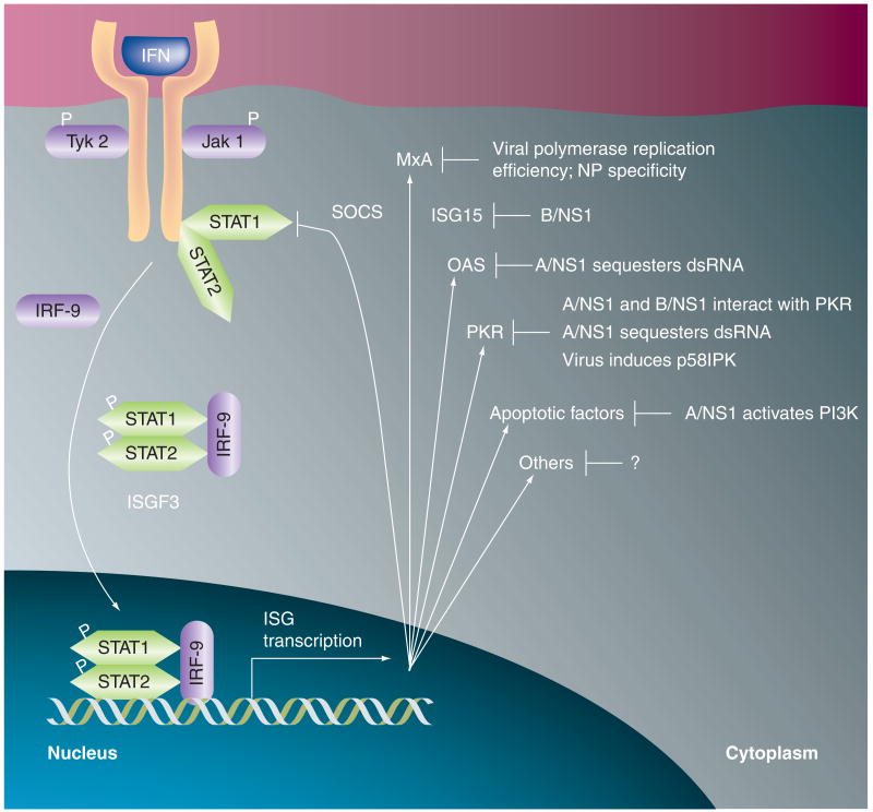
Influenza viruses are globally important human respiratory pathogens. These viruses cause seasonal epidemics and occasional worldwide pandemics, both of which can vary significantly in disease severity. The virulence of a particular influenza virus strain is partly determined by its success in circumventing the host immune response. This article briefly reviews the innate mechanisms that host cells have evolved to resist virus infection, and outlines the plethora of strategies that influenza viruses have developed in order to counteract such powerful defences. The molecular details of this virus-host interplay are summarized, and the ways in which research in this area is being applied to the rational design of protective vaccines and novel antivirals are discussed.
Figures
Similar articles
-
Modulation of Innate Immune Responses by the Influenza A NS1 and PA-X Proteins.Viruses. 2018 Dec 12;10(12):708. doi: 10.3390/v10120708. Viruses. 2018. PMID: 30545063 Free PMC article. Review.
-
Immune responses to influenza virus infection.Virus Res. 2011 Dec;162(1-2):19-30. doi: 10.1016/j.virusres.2011.09.022. Epub 2011 Sep 22. Virus Res. 2011. PMID: 21963677 Review.
-
Induction and evasion of type I interferon responses by influenza viruses.Virus Res. 2011 Dec;162(1-2):12-8. doi: 10.1016/j.virusres.2011.10.017. Epub 2011 Oct 21. Virus Res. 2011. PMID: 22027189 Free PMC article. Review.
-
Evasion mechanisms of the type I interferons responses by influenza A virus.Crit Rev Microbiol. 2020 Aug;46(4):420-432. doi: 10.1080/1040841X.2020.1794791. Epub 2020 Jul 25. Crit Rev Microbiol. 2020. PMID: 32715811 Review.
-
Vaccination strategies for an influenza pandemic.J Infect Dis. 2005 Apr 15;191(8):1207-9. doi: 10.1086/428952. Epub 2005 Mar 14. J Infect Dis. 2005. PMID: 15776363 No abstract available.
Cited by
-
Avian influenza virus H5N1 induces rapid interferon-beta production but shows more potent inhibition to retinoic acid-inducible gene I expression than H1N1 in vitro.Virol J. 2012 Aug 3;9:145. doi: 10.1186/1743-422X-9-145. Virol J. 2012. PMID: 22862800 Free PMC article.
-
Engineering potent live attenuated coronavirus vaccines by targeted inactivation of the immune evasive viral deubiquitinase.Nat Commun. 2023 Feb 28;14(1):1141. doi: 10.1038/s41467-023-36754-z. Nat Commun. 2023. PMID: 36854765 Free PMC article.
-
Cytokine production in whole-blood cultures following immunization with an influenza vaccine.Hum Vaccin Immunother. 2018;14(12):2990-2998. doi: 10.1080/21645515.2018.1498435. Epub 2018 Aug 28. Hum Vaccin Immunother. 2018. PMID: 30036123 Free PMC article.
-
Comparative Analysis of Whole-Transcriptome RNA Expression in MDCK Cells Infected With the H3N2 and H5N1 Canine Influenza Viruses.Front Cell Infect Microbiol. 2019 Mar 26;9:76. doi: 10.3389/fcimb.2019.00076. eCollection 2019. Front Cell Infect Microbiol. 2019. PMID: 30972307 Free PMC article.
-
Discovery of a Novel Tetrapeptide against Influenza A Virus: Rational Design, Synthesis, Bioactivity Evaluation and Computational Studies.Pharmaceuticals (Basel). 2021 Sep 23;14(10):959. doi: 10.3390/ph14100959. Pharmaceuticals (Basel). 2021. PMID: 34681184 Free PMC article.
References
Bibliography
-
- Palese P, Shaw ML. Orthomyxoviridae: the viruses and their replication. In: Knipe DM, Howley PM, editors. Fields Virology. Lippincott Williams & Wilkins; PA, USA: 2007. pp. 1647–1689.
-
- Klenk HD, Cox NJ, Lamb RA, et al. Orthomyxoviridae. In: Büchen-Osmond C, editor. ICTVdB – The Universal Virus Database. Columbia University; NY, USA: 2004.
-
- Infectious Diseases Society of America (IDSA) Avian Influenza (Bird Flu): Implications for Human Disease. 2007
-
- Wright PF, Webster RG. Orthomyxoviruses. In: Knipe DM, Howley PM, editors. Fields Virology. Lippincott Williams & Wilkins; PA, USA: 2007. pp. 1533–1579.
Website
-
- WHO. Influenza fact sheet 211. 2009. www.who.int/mediacentre/factsheets/fs211/en/
Publication types
MeSH terms
Substances
Grants and funding
LinkOut - more resources
Full Text Sources
Other Literature Sources
Medical