

Pancreatic adenocarcinoma in a patient with situs inversus: a case report of this rare coincidence
- PMID: 20021643
- PMCID: PMC2803176
- DOI: 10.1186/1477-7819-7-98
Pancreatic adenocarcinoma in a patient with situs inversus: a case report of this rare coincidence
Abstract
Background: Situs inversus (SI) is a relatively rare occurrence in patients with pancreatic adenocarcinoma. Pancreatic resection in these patients has rarely been described. CT scan imaging is a principle modality for detecting pancreatic cancer and its use in SI patients is seldom reported.
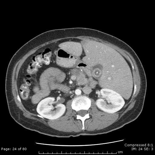
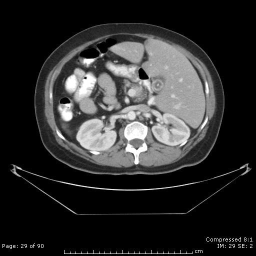
Case presentation: We report a 48 year old woman with SI who, despite normal CT scan 8 months earlier, presented with obstructive jaundice and a pancreatic head mass requiring a pancreaticoduodenectomy. The surgical pathology report demonstrated pancreatic adenocarcinoma.
Conclusion: SI is a rare condition with concurrent pancreatic cancer being even rarer. Despite the rarity, pancreaticoduodenectomy in these patients for resectable lesions is safe as long as special consideration to the anatomy is taken. Additionally, radiographic imaging has significantly improved detection of early pancreatic cancer; however, there continues to be a need for improved detection of small neoplasms.
Figures

References
-
- Legmann P, Vignaux O, Dousset B, Baraza AJ, Palazzo L, Dumontier I, Coste J, Louvel A, Roseau G, Couturier D, Bonnin A. Pancreatic tumors: comparison of dual-phase helical CT and endoscopic sonography. AJR Am J Roentgenol. 1998;170:1315–1322. - PubMed
-
- Kosaki K, Casey B. Genetics of human left-right axis malformations. Cell & Developmental Biology. 1998;9:89–99. - PubMed
Publication types
MeSH terms
LinkOut - more resources
Full Text Sources
Medical
