

Reduced expression of cenp-e in human hepatocellular carcinoma
- PMID: 20021663
- PMCID: PMC2804602
- DOI: 10.1186/1756-9966-28-156
Reduced expression of cenp-e in human hepatocellular carcinoma
Abstract
Background: CENP-E, one of spindle checkpoint proteins, plays a crucial role in the function of spindle checkpoint. Once CENP-E expression was interrupted, the chromosomes can not separate procedurally, and may result in aneuploidy which is a hallmark of most solid cancers, such as hepatocellular carcinoma (HCC). We investigate the expression of CENP-E in human hepatocellular carcinoma,. and analyze the effect of low CENP-E expression on chromosome separation in normal liver cell line (LO2).
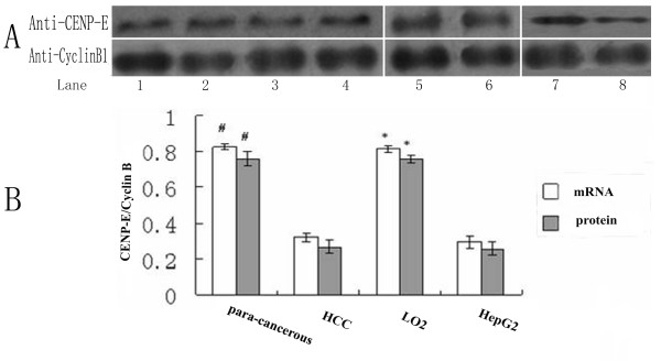
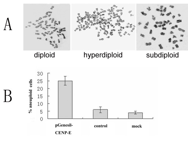
Methods: We determined its levels in HCC and para-cancerous tissues, human hepatocellular carcinoma-derived cell line (HepG2) and LO2 cell line using real time quantitative PCR (QPCR) and Western blot. Further to know whether reduction in CENP-E expression impairs chromosomes separation in LO2 cells. we knocked down CENP-E using shRNA expressing vector and then count the aneuploid in LO2 cells using chromosomal counts assay.
Results: We found that both CENP-E mRNA and protein levels were significantly reduced in HCC tissues and HepG2 cells compared with para-cancerous tissues and LO2 cells, respectively. A significantly-increased proportion of aneuploid in these down-knocked LO2 cells compared with those treated with control shRNA vector.
Conclusions: Together with other results, these results reveal that CENP-E expression was reduced in human HCC tissue, and low CENP-E expression result in aneuploidy in LO2 cells.
Figures


Similar articles
-
[Expression of centromere protein A in hepatocellular carcinoma].Zhonghua Bing Li Xue Za Zhi. 2007 Mar;36(3):175-8. Zhonghua Bing Li Xue Za Zhi. 2007. PMID: 17535684 Chinese.
-
Hepatitis B virus X protein mutant upregulates CENP-A expression in hepatoma cells.Oncol Rep. 2012 Jan;27(1):168-73. doi: 10.3892/or.2011.1478. Epub 2011 Sep 28. Oncol Rep. 2012. PMID: 21956590
-
Overexpression of CENP-H as a novel prognostic biomarker for human hepatocellular carcinoma progression and patient survival.Oncol Rep. 2013 Nov;30(5):2238-44. doi: 10.3892/or.2013.2675. Epub 2013 Aug 20. Oncol Rep. 2013. PMID: 23970101
-
Reduced expression of CENP-E contributes to the development of hepatocellular carcinoma and is associated with adverse clinical features.Biomed Pharmacother. 2020 Mar;123:109795. doi: 10.1016/j.biopha.2019.109795. Epub 2019 Dec 24. Biomed Pharmacother. 2020. PMID: 31881483
-
[The expression and clinopathological significance of miR-130b in human hepatocellular carcinoma].Xi Bao Yu Fen Zi Mian Yi Xue Za Zhi. 2016 Mar;32(3):387-92. Xi Bao Yu Fen Zi Mian Yi Xue Za Zhi. 2016. PMID: 26927562 Chinese.
Cited by
-
Deregulation of the spindle assembly checkpoint is associated with paclitaxel resistance in ovarian cancer.J Ovarian Res. 2018 Apr 4;11(1):27. doi: 10.1186/s13048-018-0399-7. J Ovarian Res. 2018. PMID: 29618387 Free PMC article.
-
Processing of kansui roots stir-baked with vinegar reduces kansui-induced hepatocyte cytotoxicity by decreasing the contents of toxic terpenoids and regulating the cell apoptosis pathway.Molecules. 2014 Jun 3;19(6):7237-54. doi: 10.3390/molecules19067237. Molecules. 2014. PMID: 24896263 Free PMC article.
-
Recent Trends of microRNA Significance in Pediatric Population Glioblastoma and Current Knowledge of Micro RNA Function in Glioblastoma Multiforme.Int J Mol Sci. 2020 Apr 27;21(9):3046. doi: 10.3390/ijms21093046. Int J Mol Sci. 2020. PMID: 32349263 Free PMC article. Review.
-
Highly expressed centromere protein L indicates adverse survival and associates with immune infiltration in hepatocellular carcinoma.Aging (Albany NY). 2021 Oct 4;13(19):22802-22829. doi: 10.18632/aging.203574. Epub 2021 Oct 4. Aging (Albany NY). 2021. PMID: 34607313 Free PMC article.
-
Role of motor proteins in human cancers.Saudi J Biol Sci. 2022 Dec;29(12):103436. doi: 10.1016/j.sjbs.2022.103436. Epub 2022 Sep 6. Saudi J Biol Sci. 2022. PMID: 36131778 Free PMC article. Review.
References
MeSH terms
Substances
LinkOut - more resources
Full Text Sources
Medical
