. 2009 Dec 18:7:109.
doi: 10.1186/1479-5876-7-109.

Short-term cultured, interleukin-15 differentiated dendritic cells have potent immunostimulatory properties

Affiliations

Short-term cultured, interleukin-15 differentiated dendritic cells have potent immunostimulatory properties

Sébastien Anguille et al. J Transl Med. .

Abstract

Background: Optimization of the current dendritic cell (DC) culture protocol in order to promote the therapeutic efficacy of DC-based immunotherapy is warranted. Alternative differentiation of monocyte-derived DCs using granulocyte macrophage colony-stimulating factor (GM-CSF) and interleukin (IL)-15 has been propagated as an attractive strategy in that regard. The applicability of these so-called IL-15 DCs has not yet been firmly established. We therefore developed a novel pre-clinical approach for the generation of IL-15 DCs with potent immunostimulatory properties.

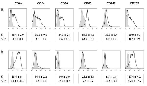
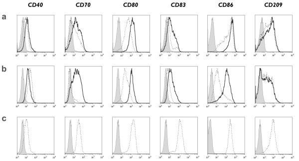
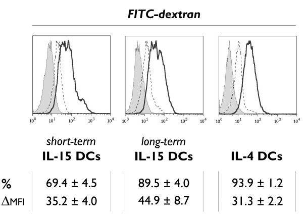
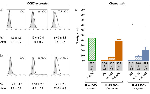
Methods: Human CD14+ monocytes were differentiated with GM-CSF and IL-15 into immature DCs. Monocyte-derived DCs, conventionally differentiated in the presence of GM-CSF and IL-4, served as control. Subsequent maturation of IL-15 DCs was induced using two clinical grade maturation protocols: (i) a classic combination of pro-inflammatory cytokines (tumor necrosis factor-alpha, IL-1beta, IL-6, prostaglandin E2) and (ii) a Toll-like receptor (TLR)7/8 agonist-based cocktail (R-848, interferon-gamma, TNF-alpha and prostaglandin E2). In addition, both short-term (2-3 days) and long-term (6-7 days) DC culture protocols were compared. The different DC populations were characterized with respect to their phenotypic profile, migratory properties, cytokine production and T cell stimulation capacity.

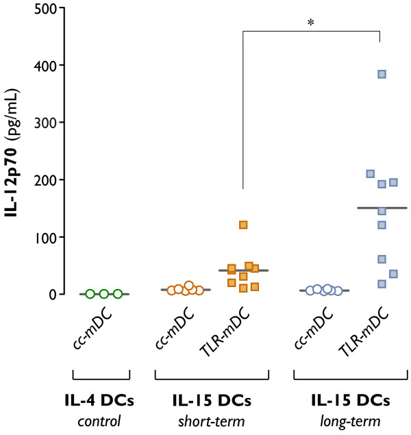
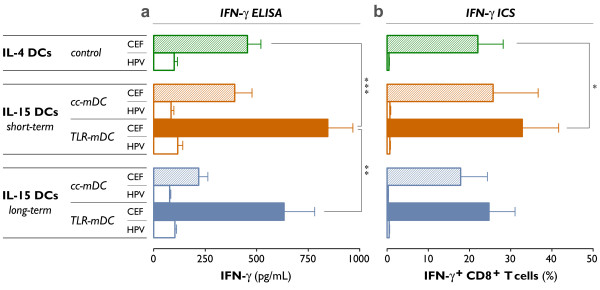
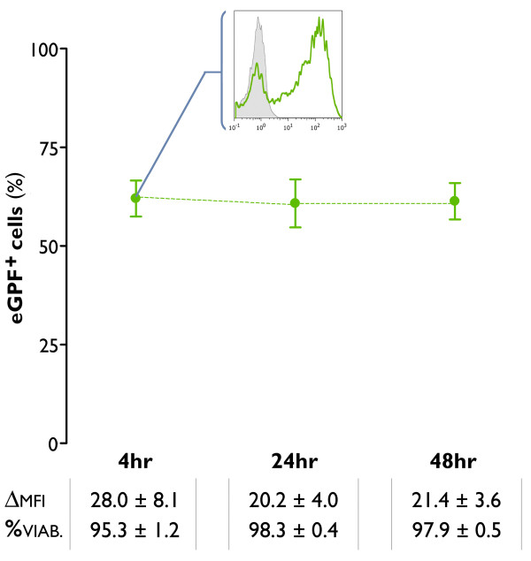
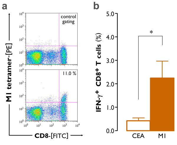
Results: The use of a TLR7/8 agonist-based cocktail resulted in a more optimal maturation of IL-15 DCs, as reflected by the higher phenotypic expression of CD83 and costimulatory molecules (CD70, CD80, CD86). The functional superiority of TLR7/8-activated IL-15 DCs over conventionally matured IL-15 DCs was evidenced by their (i) higher migratory potential, (ii) advantageous cytokine secretion profile (interferon-gamma, IL-12p70) and (iii) superior capacity to stimulate autologous, antigen-specific T cell responses after passive peptide pulsing. Aside from a less pronounced production of bioactive IL-12p70, short-term versus long-term culture of TLR7/8-activated IL-15 DCs resulted in a migratory profile and T cell stimulation capacity that was in favour of short-term DC culture. In addition, we demonstrate that mRNA electroporation serves as an efficient antigen loading strategy of IL-15 DCs.

Conclusions: Here we show that short-term cultured and TLR7/8-activated IL-15 DCs fulfill all pre-clinical prerequisites of immunostimulatory DCs. The results of the present study might pave the way for the implementation of IL-15 DCs in immunotherapy protocols.


Figures

Figure 1
Figure 1
Phenotypic characteristics of immature IL-15 DCs. Immature DCs were analyzed by flow cytometry for the expression of CD1a, CD14, CD56, CD80, CD207 (Langerin) and CD209 (DC-SIGN). The histograms represent the expression of the indicated cell surface antigens (bold-line histograms) and the corresponding isotype controls (grey-filled histograms). The mean ± SEM percentage of positive cells (%) and delta MFI ± SEM (ΔMFI) were calculated as specified in the "Methods" section (n = 3-6). (a) Phenotype of monocyte-derived DCs generated in the presence of GM-CSF + IL-15 and harvested at the immature stage 2-3 days after initiation of the DC culture. (b) Corresponding phenotypic profile of conventional immature DCs, differentiated in the presence of GM-CSF + IL-4.
Figure 2
Figure 2
Phenotypic characteristics of mature IL-15 DCs. Immunophenotypic expression of CD40, CD70, CD80, CD83, CD86 and CD209 (DC-SIGN) by (a) short-term cultured IL-15 DCs, (b) long-term cultured IL-15 DCs and (c) conventional IL-4 DCs after maturation induction with either a classic maturation cocktail (cc-mDC; dashed-line histograms) or a TLR7/8 ligand-containing mixture (TLR-mDC; bold-line histograms). Isotype controls are represented by the grey-filled histograms. A detailed overview of the flow cytometry data of mature DCs is provided in "Additional file 1" (n = 4).
Figure 3
Figure 3
Mannose receptor-mediated endocytosis of FITC-dextran particles. Histogram overlays depicting the in vitro uptake of FITC-dextran molecules by immature DCs, respectively short-term cultured IL-15 DCs (left), long-term cultured IL-15 DCs (middle) and control IL-4 DCs (right). The FITC-dextran endocytosis at 37°C (bold-line histograms) is compared to the non-specific fluorescence at 4°C (dashed-line histograms) and to the autofluorescence from unlabeled samples (grey-filled histograms), as described in "Methods". The uptake of FITC-dextran was quantified as mean ± SEM percentage of FITC-dextran positive cells (%) and as delta MFI ± SEM (ΔMFI), which was calculated by subtracting the MFI value of the non-specific FITC-dextran uptake at 4°C from the MFI value obtained at 37°C (n = 3).
Figure 4
Figure 4
CCR7 expression and migratory capacity. Histogram overlays comparing the CCR7 expression on (a) short-term cultured and (b) long-term cultured IL-15 DCs, either at the immature stage (iDC) or at the mature stage (cc-mDC: + TNF-α, IL1β, IL-6 and PGE2 for the last 24 hours; TLR-mDC: + R-848, IFN-γ, TNF-α and PGE2 for the last 24 hours). Bold-line histograms represent the CCR7-specific staining, whereas the corresponding isotype controls are indicated by grey-filled histograms (n = 3). (c) Migration of the indicated DC subsets towards CCL21 in a Transwell chemotaxis assay. The mean ± SEM percentages of migrated cells after 180 min were calculated according to the formula specified in "Methods" (n = 3-6; *, P = 0.01). The values shown in the grey bars represent the cell viabilities of the different DC subsets (mean ± SEM; n = 3-6).
Figure 5
Figure 5
IL-12p70 production following CD40 ligation ("signal-3 assay"). Dendritic cells were differentiated in the presence of GM-CSF + IL-4 for 6 days (control IL-4 DCs), or in the presence of GM-CSF + IL-15 for 2 days (short-term IL-15 DCs) or 6 days (long-term IL-15 DCs). Dendritic cell maturation was induced by addition of two different maturation cocktails 24 hr prior to DC harvest (cc-mDC: TNF-α, IL1β, IL-6 and PGE2; TLR-mDC: R-848, IFN-γ, TNF-α and PGE2). Production of the Th1-polarizing cytokine IL-12p70 was assessed by ELISA after a 24-hr coculture of mDCs and CD40L-expressing 3T3 fibroblasts, as specified in "Methods". Results are shown from 3-9 independent experiments, each symbol expressing the mean of triplicate ELISA values obtained from one individual donor. The horizontal bars represent the mean IL-12p70 production in pg/mL per condition (*, P = 0.02).
Figure 6
Figure 6
Induction of viral antigen-specific CD8+ T cell responses. As described previously, short-term and long-term cultured IL-15 DCs were matured using two different maturation cocktails (cc-mDC: TNF-α, IL1β, IL-6 and PGE2; TLR-mDC: R-848, IFN-γ, TNF-α and PGE2). Conventionally matured IL-4 DCs were used as a control (control IL-4 DCs). The mDCs were harvested, pulsed with a pool of cytomegalovirus-, Epstein-Barr virus- and influenza a virus (CEF)-derived peptides, and cocultured with autologous PBLs for 7 days. Viral antigen-specific CD8+ T cell responses were determined after this 7-day period and a short restimulation with the CEF peptide pool (CEF; filled bars). As specified in "Methods", the antigen-specific production of IFN-γ was assessed using two techniques: (a) ELISA to detect the amount of IFN-γ produced after restimulation (pg/mL) and (b) ICS to determine the % of IFN-γ+ CD8+ T cells. The non-specific IFN-γ release in response to restimulation with an irrelevant HPV peptide pool is shown (HPV; unfilled bars). Results are expressed as mean ± SEM of three independent experiments (*, P = 0.03; **, P = 0.006; ***, P < 0.001).
Figure 7
Figure 7
mRNA transfectability of mature IL-15 DCs. Monocytes were cultured for 2 days with GM-CSF + IL-15, followed by a 24-hr incubation with a TLR7/8 agonist-based maturation cocktail (TLR-mDC). The resultant mDCs were harvested and electroporated with mRNA encoding the enhanced green fluorescent protein (eGFP). The green dots represent the mean ± SEM percentages of eGFP+ cells, as assessed by flow cytometry at different time points post-electroporation (4 hr, 24 hr, 48 hr). The insert shows a representative histogram overlay in which the flow cytometric eGFP expression 4 hr post-electroporation (green line histogram) is compared with the expression in a mock-electroporated negative control (grey-filled histogram). The values below indicate the delta MFI ± SEM of the eGFP expression (ΔMFI) and the mean ± SEM percentage of viable cells (%) at 4 hr, 24 hr and 48 hr following mRNA electrotransfection of IL-15 DCs (n = 5).
Figure 8
Figure 8
Induction of antigen-specific CD8+ T cell responses by mRNA-electroporated mature IL-15 DCs. Short-term cultured IL-15 DCs were matured with our TLR7/8 agonist-based maturation cocktail (TLR-mDC), electroporated with mRNA encoding the influenza virus matrix protein M1 and cocultured with autologous PBLs for 6 days. (a) The expansion of M1-tetramer binding CD8+ T cells was determined by flow cytometry. The lower dot plot represents the observed percentage of M1-tetramer+ CD8+ T cells in one representative donor (n = 4; mean ± SEM percentage of M1-tetramer+ CD8+ T cells: 4.4 ± 2.9). Correct positioning of the M1-tetramer+ CD8+ gate was defined by the respective negative control, as exemplified in the upper dot plot. (b) Simultaneously, a fraction of the PBL was harvested and stimulated with an irrelevant HLA-A*0201-restricted peptide (CEA) or rechallenged with the immunodominant influenza matrix protein (M1). The mean ± SEM percentage of antigen-specific IFN-γ+ CD8+ T cells was determined by ICS, as specified in the "Methods" section (n = 4; *, P = 0.03).

Similar articles

Cited by

References

    1. Steinman RM, Cohn ZA. Identification of a novel cell type in peripheral lymphoid organs of mice. II. Functional properties in vitro. J Exp Med. 1974;139:380–397. doi: 10.1084/jem.139.2.380. - DOI - PMC - PubMed
    1. Steinman RM, Cohn ZA. Identification of a novel cell type in peripheral lymphoid organs of mice. I. Morphology, quantitation, tissue distribution. J Exp Med. 1973;137:1142–1162. doi: 10.1084/jem.137.5.1142. - DOI - PMC - PubMed
    1. Steinman RM, Banchereau J. Taking dendritic cells into medicine. Nature. 2007;449:419–426. doi: 10.1038/nature06175. - DOI - PubMed
    1. Palucka AK, Ueno H, Fay JW, Banchereau J. Taming cancer by inducing immunity via dendritic cells. Immunol Rev. 2007;220:129–150. doi: 10.1111/j.1600-065X.2007.00575.x. - DOI - PubMed
    1. Tuyaerts S, Aerts JL, Corthals J, Neyns B, Heirman C, Breckpot K, Thielemans K, Bonehill A. Current approaches in dendritic cell generation and future implications for cancer immunotherapy. Cancer Immunol Immunother. 2007;56:1513–1537. doi: 10.1007/s00262-007-0334-z. - DOI - PMC - PubMed

Publication types

MeSH terms