. 2009 Dec 19:6:38.
doi: 10.1186/1742-2094-6-38.

Cellular injury and neuroinflammation in children with chronic intractable epilepsy

Affiliations

Cellular injury and neuroinflammation in children with chronic intractable epilepsy

Jieun Choi et al. J Neuroinflammation. .

Abstract

Objective: To elucidate the presence and potential involvement of brain inflammation and cell death in neurological morbidity and intractable seizures in childhood epilepsy, we quantified cell death, astrocyte proliferation, microglial activation and cytokine release in brain tissue from patients who underwent epilepsy surgery.

Methods: Cortical tissue was collected from thirteen patients with intractable epilepsy due to focal cortical dysplasia (6), encephalomalacia (5), Rasmussen's encephalitis (1) or mesial temporal lobe epilepsy (1). Sections were processed for immunohistochemistry using markers for neuron, astrocyte, microglia or cellular injury. Cytokine assay was performed on frozen cortices. Controls were autopsy brains from eight patients without history of neurological diseases.

Results: Marked activation of microglia and astrocytes and diffuse cell death were observed in epileptogenic tissue. Numerous fibrillary astrocytes and their processes covered the entire cortex and converged on to blood vessels, neurons and microglia. An overwhelming number of neurons and astrocytes showed DNA fragmentation and its magnitude significantly correlated with seizure frequency. Majority of our patients with abundant cell death in the cortex have mental retardation. IL-1beta, IL-8, IL-12p70 and MIP-1beta were significantly increased in the epileptogenic cortex; IL-6 and MCP-1 were significantly higher in patients with family history of epilepsy.

Conclusions: Our results suggest that active neuroinflammation and marked cellular injury occur in pediatric epilepsy and may play a common pathogenic role or consequences in childhood epilepsy of diverse etiologies. Our findings support the concept that immunomodulation targeting activated microglia and astrocytes may be a novel therapeutic strategy to reduce neurological morbidity and prevent intractable epilepsy.


Figures

Figure 1
Figure 1
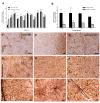
Quantification of CD68 immunoreactive microglia in the cortex (cortical GM) and white matter (subcortical WM). (A) CD68-immunoreactivity in individual patients (patients No. 1 to 11) and controls (n = 5). Significant microglial activation was noted in 8/11 patients (73%, p < 0.05). (B) Fold changes in the CD68-immunoreactivity in epilepsy subgroups compared to controls. Note the magnitude of microglia activation (nearly 20-fold increase in the cortex) in FCD and EM is comparable to RE. (C-J) CD68-immunoreactiviy in the cortex (upper panel) and white matter (lower panel). Controls show very rare microglia (C, G). Focal cortical dysplasia (D, H) and encephalomalacia (E, I) show uniform increase in microglia while Rasmussen's encephalitis (F, J) show microglial aggregates. Bar = 20 μm. *p < 0.05, ** p < 0.01, *** p < 0.001, one-way ANOVA.
Figure 2
Figure 2
Quantification of GFAP immunoreactive astrocytes in the cortex and white matter. (A) GFAP-immunoreactivity in individual patients (patients No. 1 to 11) and controls (n = 5). (B) Fold changes in the GFAP-immunoreactivity in epilepsy subgroups compared to controls. (C, D & E) Control: cortex, gray-white junction and white matter. (F, G & H) Epilepsy case: cortex, gray-white junction and white matter. Notice loss of clear gray-white demarcation in epilepsy case (G) compared to control (D). (I-K) High magnification of an epilepsy case. (I) Expanded subpial gliosis. (J) Cortical astrocytes with numerous thin and elongated processes, morphologically resembling fibrillary astrocytes. (K) Astrocytic end feet adhering to the wall of blood vessel. Bars: C-H = 100 μm; I-K = 20 μm. * p < 0.05, ** p < 0.01, *** p < 0.001 by one-way ANOVA.
Figure 3
Figure 3
Triple immunofluorescence confocal images of neurons (green), microglia (blue) and astrocytes (red). Low magnification view of cortex (A) and white matter (B). (C--H) Higher magnification view showing close contact between microglia and astrocytes (C, D), neurons and microglia (E, F) and neurons and astrocytes (G, H). Activated microglia tend to co-localize with astrocytes (p = 0.054) (I). Activated microglia is inversely correlated with DNA fragmentation (p < 0.02, r = -0.38) (J). Bar = 20 μ m.
Figure 4
Figure 4
Quantification and Triple immunofluorescence confocal images of DNA fragmentation in the cortex and white matter. (A) DNA fragmentation in individual patients (patients No. 1 to 11) and controls (n = 5). Notice that the majority of our patients [6/10, except RE (No. 11)] showed diffuse cell injury that is comparable to the patient with Rasmussen's encephalitis (No. 11). (B) Fold changes in DNA fragmentation in epilepsy subgroups over controls. (C) The magnitude of DNA fragmentation is significantly correlated with seizure frequency prior to surgery (p = 0.002). (D) Low magnification view of a control shows no evidence of DNA fragmentation. (E-G) Epilepsy case. Low and high magnification of cortex (F) and white matter (G) show increased DNA fragmentations. (H-K) Higher magnification view of DNA fragmentation in neurons (black arrows), oligodendrocytes (green arrows) and astrocytes (blue arrows) in cortex (H, I and J) and white matter (K). Microglia were spared (red arrows). The identification of different cell types was made based on nuclear morphology. Neurons have large vesicular nuclei and oligodendrocytes have round uniform nuclei while astrocytes have bipolar elongated nuclei with irregular borders. Small rod-shaped nuclei of microglia show only faint staining. (L-O) Fluoro-Jade B staining in control (L) and epilepsy cases (M-O). Positive staining in the cortex (M, N) and white matter (O) of epilepsy cases and not in controls are consistent with cell injury noted by in situ end labeling of DNA fragmentation. (P-S) Triple immunofluorescence confocal images of cellular subtypes showing DNA fragmentation in the cortex (P, R) and white matter (Q, S) of focal cortical dysplasia patient; Low magnification (P) shows that neurons comprise the majority of cells with DNA fragmentation. Double labeling of neuron and DNA fragmentation is magnified and microglia closely associated with these neurons are also labeled with fragmented DNA (Fig. R4). DNA fragmentation (blue); microglia (red); neuron (green). (P, R) White matter. DNA fragmentation (blue); astrocyte (red); neuron (green). Low (Q) and high magnification (S) show double labeling of astrocytes with DNA fragmentation. Bars: D-G = 100 μ m; H-S = 20 μ m. * p < 0.05, ** p < 0.01, *** p < 0.001, one-way ANOVA.
Figure 5
Figure 5
Cytokine analysis by ELISA. IL-1β (A), IL-8 (B) and MIP-1β (C) are significantly increased in brains of patients with epilepsy (p < 0.05) whereas. IL-6 (D), MCP-1 (E) and IP-10 (F) show sporadic and variable increases in our patients with epilepsy. Note that some of our patients show highly elevated cytokine release comparable to Rasmussen's encephalitis (black circle). IL-6 (G) and MCP-1 (H) are significantly increased in brains from patients with a family history of epilepsy (p < 0.05). Rasmussen's encephalitis patient (black circle) is the only one who shows high IL-6 and MCP-1 levels among the patients without family history of epilepsy.

Similar articles

Cited by

References

    1. Allan SM, Rothwell NJ. Cytokines and acute neurodegeneration. Nat Rev Neurosci. 2001;2:734–744. doi: 10.1038/35094583. - DOI - PubMed
    1. Vezzani A, Granata T. Brain inflammation in epilepsy: experimental and clinical evidence. Epilepsia. 2005;46:1724–1743. doi: 10.1111/j.1528-1167.2005.00298.x. - DOI - PubMed
    1. Ravizza T, Gagliardi B, Noe F, Boer K, Aronica E, Vezzani A. Innate and adaptive immunity during epileptogenesis and spontaneous seizures: Evidence from experimental models and human temporal lobe epilepsy. Neurobiol Dis. 2008;29:142–160. doi: 10.1016/j.nbd.2007.08.012. - DOI - PubMed
    1. Crespel A, Coubes P, Rousset MC, Brana C, Rougier A, Rondouin G, Bockaert J, Baldy-Moulinier M, Lerner-Natoli M. Inflammatory reactions in human medial temporal lobe epilepsy with hippocampal sclerosis. Brain Res. 2002;952:159–169. doi: 10.1016/S0006-8993(02)03050-0. - DOI - PubMed
    1. Boer K, Spliet WG, van Rijen PC, Redeker S, Troost D, Aronica E. Evidence of activated microglia in focal cortical dysplasia. J Neuroimmunol. 2006;173:188–195. doi: 10.1016/j.jneuroim.2006.01.002. - DOI - PubMed

Publication types

MeSH terms