

The longitudinal changes of BOLD response and cerebral hemodynamics from acute to subacute stroke. A fMRI and TCD study
- PMID: 20021696
- PMCID: PMC2805667
- DOI: 10.1186/1471-2202-10-151
The longitudinal changes of BOLD response and cerebral hemodynamics from acute to subacute stroke. A fMRI and TCD study
Abstract
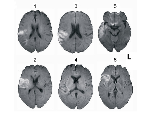
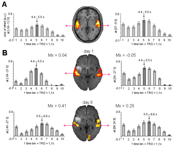
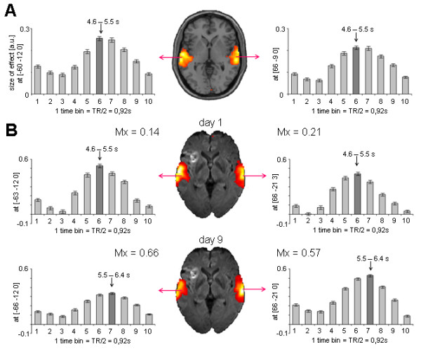
Background: By mapping the dynamics of brain reorganization, functional magnetic resonance imaging MRI (fMRI) has allowed for significant progress in understanding cerebral plasticity phenomena after a stroke. However, cerebro-vascular diseases can affect blood oxygen level dependent (BOLD) signal. Cerebral autoregulation is a primary function of cerebral hemodynamics, which allows to maintain a relatively constant blood flow despite changes in arterial blood pressure and perfusion pressure. Cerebral autoregulation is reported to become less effective in the early phases post-stroke. This study investigated whether any impairment of cerebral hemodynamics that occurs during the acute and the subacute phases of ischemic stroke is related to changes in BOLD response. We enrolled six aphasic patients affected by acute stroke. All patients underwent a Transcranial Doppler to assess cerebral autoregulation (Mx index) and fMRI to evaluate the amplitude and the peak latency (time to peak-TTP) of BOLD response in the acute (i.e., within four days of stroke occurrence) and the subacute (i.e., between five and twelve days after stroke onset) stroke phases.
Results: As patients advanced from the acute to subacute stroke phase, the affected hemisphere presented a BOLD TTP increase (p = 0.04) and a deterioration of cerebral autoregulation (Mx index increase, p = 0.046). A similar but not significant trend was observed also in the unaffected hemisphere. When the two hemispheres were grouped together, BOLD TTP delay was significantly related to worsening cerebral autoregulation (Mx index increase) (Spearman's rho = 0.734; p = 0.01).
Conclusions: The hemodynamic response function subtending BOLD signal may present a delay in peak latency that arises as patients advance from the acute to the subacute stroke phase. This delay is related to the deterioration of cerebral hemodynamics. These findings suggest that remodeling the fMRI hemodynamic response function in the different phases of stroke may optimize the detection of BOLD signal changes.
Figures
References
-
- Rossini PM, Altamura C, Ferreri F, Melgari JM, Tecchio F, Tombini M, Pasqualetti P, Vernieri F. Neuroimaging experimental studies on brain plasticity in recovery from stroke. Eura Medicophys. 2007;43:241–54. - PubMed
-
- Ward NS. Future perspectives in functional neuroimaging in stroke recovery. Eura Medicophys. 2007;43:285–94. - PubMed
MeSH terms
Substances
LinkOut - more resources
Full Text Sources
Medical
Research Materials
