. 2010 Mar-Apr;24(2):110-20.
doi: 10.2500/ajra.2010.24.3435.

Neuropathology of the olfactory mucosa in chronic rhinosinusitis

Affiliations

Neuropathology of the olfactory mucosa in chronic rhinosinusitis

Karen K Yee et al. Am J Rhinol Allergy. 2010 Mar-Apr.

Abstract

Background: Chronic rhinosinusitis (CRS) is a complex heterogeneous inflammatory disease that affects the nasal cavity, but the pathological examination of the olfactory mucosa (OM) in this disease has been limited.

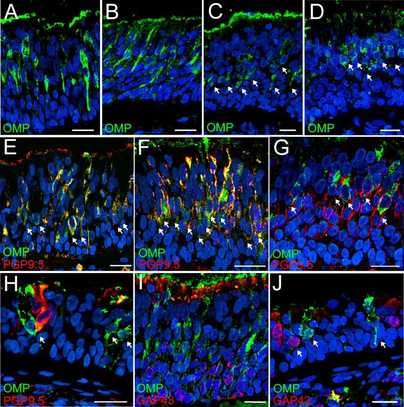
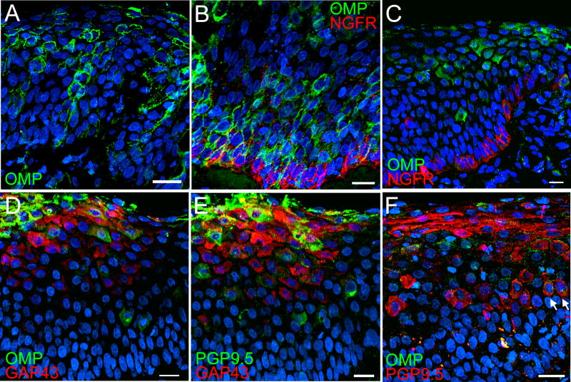
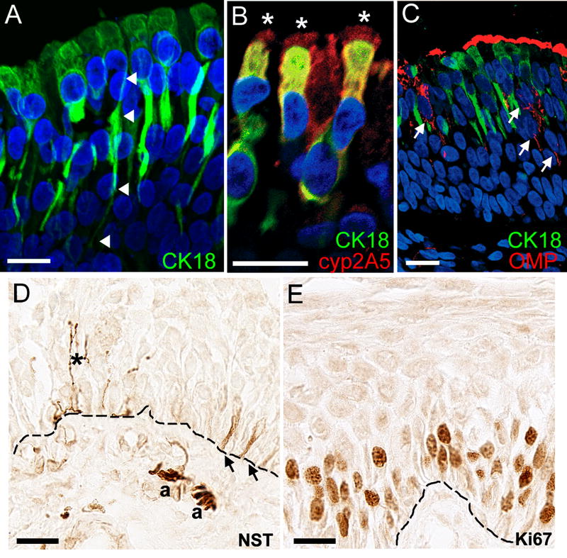
Methods: Nasal biopsy specimens were obtained from 20 control subjects and 50 CRS patients in conjunction with clinical assessments. Histopathology of these nasal biopsy specimens was performed and immunohistochemistry was used to characterize nonneuronal, neuronal, and inflammatory cells in the OM. These OM characteristics were then evaluated to determine the degree to which pathological features may be related to smell loss in CRS.

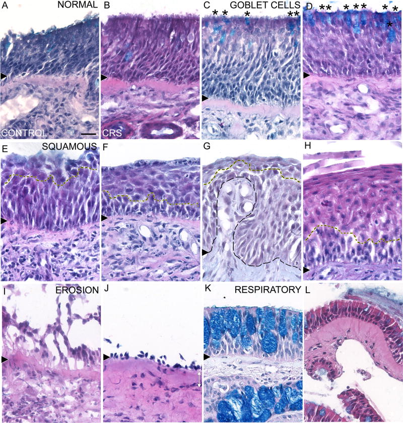
Results: Histopathological examination of control and CRS OM revealed changes in the normal pseudostratified olfactory epithelium (OE): intermixing of goblet cells, metaplasia to squamous-like cells, and erosion of the OE. Lower percentages of normal epithelium and olfactory sensory neurons were found in CRS OE compared with controls. Relative to other CRS patients, those with anosmia had the greatest amount of OE erosion, the highest density of eosinophils infiltrating the OE, and exhibited the most extensive abnormalities on CT and endoscopic examination, including being significantly more likely to exhibit nasal polyposis.

Conclusion: Our results suggest that OM pathology observed in nasal biopsy specimens can assist in understanding the degree of epithelial change and sensorineural damage in CRS and the potential for olfactory loss.


Conflict of interest statement

We have no conflict of interest nor have financial disclosure to report.

Figures

Figure 1
Figure 1. Epithelial Morphologies of Control and CRS Nasal Biopsies
A – B. Normal pseudostratified sensory epithelium was present in control (A) and CRS biopsies (B). C – D. Intermixing of Alcian Blue-stained goblet cells (asterisks) in normal pseudostratified epithelium was observed in the control (C) and CRS biopsies (D). E – H. Examples of varying degrees of epithelial transformation into squamous layers. Mild squamous metaplasia with a few layers of cuboid-shaped squamous-like cells (yellow dashed line, E) and moderate squamous metaplasia with a thicker layer of distinctive squamous-like cells (yellow dashed line, F) were observed in control biopsies. Invaginations of lamina propria (black dashed line) into mild squamous metaplasia (yellow dashed line) were often observed in CRS biopsies (G). More severe squamous metaplasia composed of several layers of distinctive squamous-like cells and flattened cells at the apical surface (yellow dashed line) was also frequently observed in CRS biopsies (H). I – J. Reduction in or loss of epithelial integrity led to erosion of cellular layers in both control (I) and CRS biopsies (J). K – L. Respiratory epithelium was identified either by a high density of Alcian Blue-stained goblet cells in the epithelium (K) or thin epithelial layers. Note also the Alcian Blue-stained mucus cells in the lamina propria. Black arrowhead = basement membrane. Scale bar = 20µm.
Figure 2
Figure 2. Immunohistochemical Labeling of Olfactory Sensory Neurons
A–D. Bipolar mature OMP-ir OSNs (green) were found in the middle layers of the normal pseudostratified OE of control (A) and CRS biopsies (B). OMP-ir OSNs (green, arrows) were also observed in goblet/intermixed OE of control (C) and CRS biopsies (D) though with less distinguishable OMP labeled processes. To further demonstrate the presence of sensory epithelium in our nasal biopsies, the OE regions in Fig 2A–D correspond to the OE region in Fig 1A–D. E–H. Double immunofluorescence labeling with OMP and PGP9.5 found the expression of these neuronal proteins varied in mature OSNs. OMP-ir cells (green, arrows) were labeled with PGP9.5 (red), especially in the processes (E–F). In other OE region of the same control biopsy, a few OMP-ir cells with PGP9.5 labeled processes were observed among PGP-ir cells (G). In an OE region with scattered neurons, a few OMP-ir cells with no PGP9.5 labeling were observed (arrows, H). I–J. Layers of immature GAP-ir cells (red) were observed below mature OMP-ir OSNs (green) suggesting olfactory neurogenesis is still on-going in these biopsies. Blue = DAPI. Scale bar = 20µm.
Figure 3
Figure 3. Immunohistochemical Labeling of Neuronal Cells in Squamous Olfactory Epithelium
A–B. Mild squamous metaplasia disrupted the localization of OMP-ir OSNs (green) such that OSNs were observed at all cellular layers (A) and localized in layers just above NGFR-ir basal cells (red, B). The OE region in 3A corresponds to Fig 1G. C–E. In moderate squamous metaplasia, mature OMP-ir OSNs (green) become abnormally shape and scattered (C) and immature GAP43-ir cells (red) were observed with OMP-ir cells (green, D), suggesting neurogenesis is still present after squamous transformation. In adjacent sections of the same CRS biopsy, a few GAP43-ir cells (red) were also labeled weakly with PGP (green, E). F. In a biopsy with severe squamous OE, only a few abnormally shaped PGP-ir cells (red) with weak OMP-ir (green, arrows) were observed. Non-specific immunoreactivity was observed in the apical flat cells. This OE region corresponds to Fig 1H. Blue = DAPI. Scale bar = 20µm.
Figure 4
Figure 4. Immunhistochemical Evaluation of the Cellular Profile in Control and CRS Nasal Biopsies
A. CK18-ir olfactory sustentacular cells (green) were located in the apical layers of the OE. Arrowheads indicate a characteristic olfactory sustentacular cell with process extending to the basement membrane. CK18-ir was also observed in Bowman’s glands and mucus glands (data not shown). B. Cyp2A5-ir (red) was co-localized in CK18-ir olfactory sustentacular cells (green) with prominent cyp2A5-ir in the cilia (asterisks). C. The presence of OMP-ir OSNs (red) among CK18-ir olfactory sustentacular cells (green) indicates CK18-ir may assist in identifying areas of sensory epithelium. D. NST-ir was seen in trigeminal fibers extending into the OE (asterisk), cells at the basal layer (arrows), and small nerve bundles (a) in the lamina propria. E. The presence of numerous proliferating Ki67-ir cells was characteristic of squamous OE. Blue = DAPI. Basement membrane = dashed line. Scale bar = 20µm.

Similar articles

Cited by

References

    1. Cowart BJ, Young IM, Feldman RS, et al. Clinical disorders of smell and taste. Occup Med. 1997;12:465–483. - PubMed
    1. Blackwell DL, Collins JG, Coles R. Summary health statistics for U.S. adults: National Health Interview Survey, 1997. Vital Health Stat 10. 2002:1–109. - PubMed
    1. Benninger MS, Ferguson BJ, Hadley JA, et al. Adult chronic rhinosinusitis: definitions, diagnosis, epidemiology, and pathophysiology. Otolaryngol Head Neck Surg. 2003;129:S1–32. - PubMed
    1. Meltzer EO, Hamilos DL, Hadley JA, et al. Rhinosinusitis: establishing definitions for clinical research and patient care. J Allergy Clin Immunol. 2004;114:155–212. - PMC - PubMed
    1. Loury MC, Kennedy DW. Chronic Sinusitis and Nasal Polyposis. In: Getchell TV, Bartoshuk LM, Doty RL, et al., editors. Smell and Taste in Health and Disease. New York: Raven Press; 1991. pp. 517–528.

Publication types

MeSH terms