

Cholesterol depletion alters coronary artery myocyte Ca(2+) signalling in a stimulus-specific manner
- PMID: 20022108
- PMCID: PMC2824115
- DOI: 10.1016/j.ceca.2009.11.009
Cholesterol depletion alters coronary artery myocyte Ca(2+) signalling in a stimulus-specific manner
Abstract
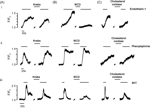
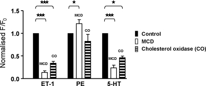
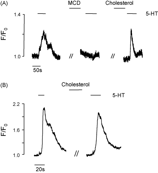
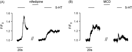
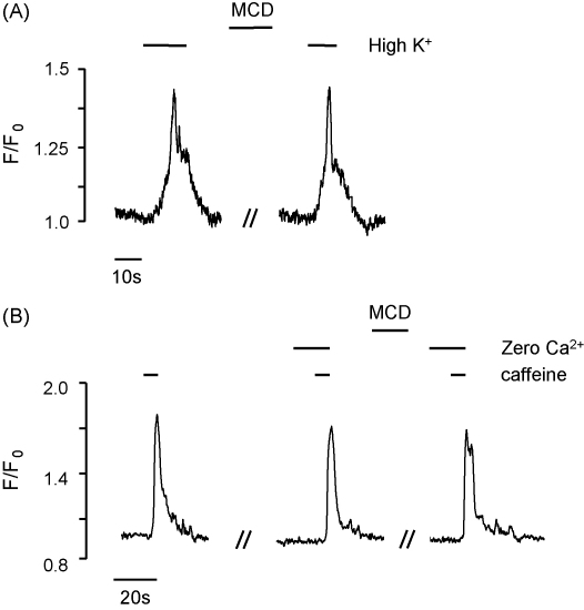
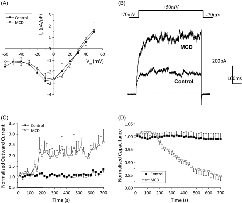
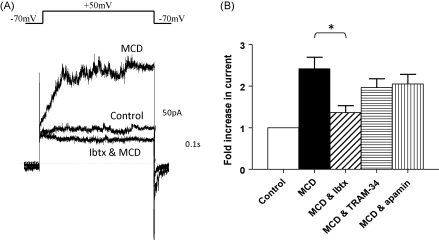
Although there is evidence that caveolae and cholesterol play an important role in myocyte signalling processes, details of the mechanisms involved remain sparse. In this paper we have studied for the first time the clinically relevant intact coronary artery and measured in situ Ca(2+) signals in individual myocytes using confocal microscopy. We have examined the effect of the cholesterol-depleting agents, methyl-cyclodextrin (MCD) and cholesterol oxidase, on high K(+), caffeine and agonist-induced Ca(2+) signals. We find that cholesterol depletion produces a stimulus-specific alteration in Ca(2+) responses; with 5-HT (10microM) and endothelin-1 (10nM) responses being selectively decreased, the phenylephrine response (100microM) increased and the responses to high K(+) (60mM) and caffeine (10mM) unaffected. Agonist-induced Ca(2+) signals were restored when cholesterol was replenished using cholesterol-saturated MCD. In additional experiments, enzymatically isolated myocytes were patch clamped. We found that cholesterol depletion caused a selective modification of ion channel function, with whole cell inward Ca(2+) current being unaltered, whereas outward K(+) current was increased, due to BK(Ca) channel activation. There was also a significant decrease in cell capacitance. These data are discussed in terms of the involvement of caveolae in receptor localisation, Ca(2+) entry pathways and SR Ca(2+) release, and the role of these in agonist signalling.
2009 Elsevier Ltd. All rights reserved.
Figures
Similar articles
-
Depletion of membrane cholesterol eliminates the Ca2+-activated component of outward potassium current and decreases membrane capacitance in rat uterine myocytes.J Physiol. 2007 Jun 1;581(Pt 2):445-56. doi: 10.1113/jphysiol.2007.129452. Epub 2007 Mar 1. J Physiol. 2007. PMID: 17331986 Free PMC article.
-
Cell culture alters Ca2+ entry pathways activated by store-depletion or hypoxia in canine pulmonary arterial smooth muscle cells.Am J Physiol Cell Physiol. 2008 Jan;294(1):C313-23. doi: 10.1152/ajpcell.00258.2007. Epub 2007 Oct 31. Am J Physiol Cell Physiol. 2008. PMID: 17977940
-
Conformation of ryanodine receptor-2 gates store-operated calcium entry in rat pulmonary arterial myocytes.Cardiovasc Res. 2016 Jul 1;111(1):94-104. doi: 10.1093/cvr/cvw067. Epub 2016 Mar 24. Cardiovasc Res. 2016. PMID: 27013634 Free PMC article.
-
Ca2+ responses of pulmonary arterial myocytes to acute hypoxia require release from ryanodine and inositol trisphosphate receptors in sarcoplasmic reticulum.Am J Physiol Lung Cell Mol Physiol. 2012 Jul;303(2):L161-8. doi: 10.1152/ajplung.00348.2011. Epub 2012 May 11. Am J Physiol Lung Cell Mol Physiol. 2012. PMID: 22582116 Free PMC article.
-
Membrane cholesterol depletion with beta-cyclodextrin impairs pressure-induced contraction and calcium signalling in isolated skeletal muscle arterioles.J Vasc Res. 2007;44(4):292-302. doi: 10.1159/000101451. Epub 2007 Mar 30. J Vasc Res. 2007. PMID: 17406121
Cited by
-
Atherosclerosis affects calcium signalling in endothelial cells from apolipoprotein E knockout mice before plaque formation.Cell Calcium. 2014 Mar;55(3):146-54. doi: 10.1016/j.ceca.2014.02.012. Epub 2014 Feb 22. Cell Calcium. 2014. PMID: 24630173 Free PMC article.
-
Atherosclerosis differentially affects calcium signalling in endothelial cells from aortic arch and thoracic aorta in Apolipoprotein E knockout mice.Physiol Rep. 2014 Oct 24;2(10):e12171. doi: 10.14814/phy2.12171. Print 2014 Oct 1. Physiol Rep. 2014. PMID: 25344475 Free PMC article.
-
Multiple cholesterol recognition/interaction amino acid consensus (CRAC) motifs in cytosolic C tail of Slo1 subunit determine cholesterol sensitivity of Ca2+- and voltage-gated K+ (BK) channels.J Biol Chem. 2012 Jun 8;287(24):20509-21. doi: 10.1074/jbc.M112.356261. Epub 2012 Apr 3. J Biol Chem. 2012. PMID: 22474334 Free PMC article.
-
The Membrane Cholesterol Modulates the Interaction Between 17-βEstradiol and the BK Channel.Front Pharmacol. 2021 Jun 11;12:687360. doi: 10.3389/fphar.2021.687360. eCollection 2021. Front Pharmacol. 2021. PMID: 34177597 Free PMC article.
-
Cholesterol activates BK channels by increasing KCNMB1 protein levels in the plasmalemma.J Biol Chem. 2021 Jan-Jun;296:100381. doi: 10.1016/j.jbc.2021.100381. Epub 2021 Feb 6. J Biol Chem. 2021. PMID: 33556372 Free PMC article.
References
-
- Brown D.A., London E. Functions of lipid rafts in biological membranes. Annu. Rev. Cell Dev. Biol. 1998;14:111–136. - PubMed
-
- Simons K., Toomre D. Lipid rafts and signal transduction. Nat. Rev. Mol. Cell Biol. 2000;1:31–39. - PubMed
-
- O’Connell K.M., Martens J.R., Tamkun M.M. Localization of ion channels to lipid Raft domains within the cardiovascular system. Trends Cardiovasc. Med. 2004;14:37–42. - PubMed
-
- Quest A.F., Leyton L., Parraga M. Caveolins, caveolae, and lipid rafts in cellular transport, signalling and disease. Biochem. Cell Biol. 2004;82:129–144. - PubMed
Publication types
MeSH terms
Substances
Grants and funding
LinkOut - more resources
Full Text Sources
Medical
Research Materials
Miscellaneous
